Calcium »
PDB 7b1l-7brv »
7bip »
Calcium in PDB 7bip: Crystal Structure of Monooxygenase RSLO1 From Streptomyces Bottropensis
Protein crystallography data
The structure of Crystal Structure of Monooxygenase RSLO1 From Streptomyces Bottropensis, PDB code: 7bip
was solved by
L.Zhang,
C.Zuo,
A.Bechthold,
O.Einsle,
with X-Ray Crystallography technique. A brief refinement statistics is given in the table below:
| Resolution Low / High (Å) | 48.53 / 1.60 |
| Space group | P 21 21 21 |
| Cell size a, b, c (Å), α, β, γ (°) | 50.912, 81.962, 160.624, 90, 90, 90 |
| R / Rfree (%) | 16.4 / 19.4 |
Calcium Binding Sites:
The binding sites of Calcium atom in the Crystal Structure of Monooxygenase RSLO1 From Streptomyces Bottropensis
(pdb code 7bip). This binding sites where shown within
5.0 Angstroms radius around Calcium atom.
In total 6 binding sites of Calcium where determined in the Crystal Structure of Monooxygenase RSLO1 From Streptomyces Bottropensis, PDB code: 7bip:
Jump to Calcium binding site number: 1; 2; 3; 4; 5; 6;
In total 6 binding sites of Calcium where determined in the Crystal Structure of Monooxygenase RSLO1 From Streptomyces Bottropensis, PDB code: 7bip:
Jump to Calcium binding site number: 1; 2; 3; 4; 5; 6;
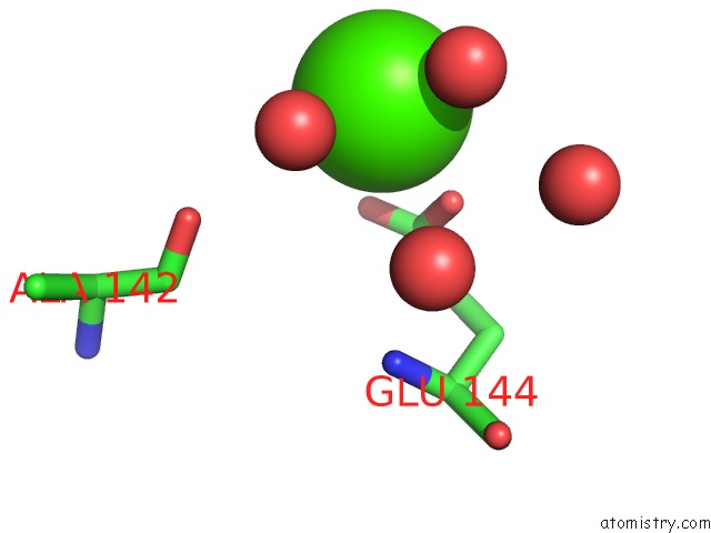
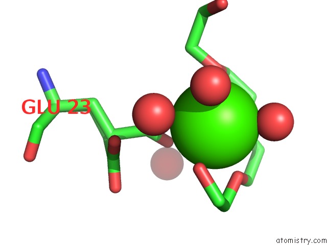
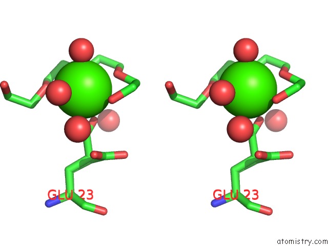
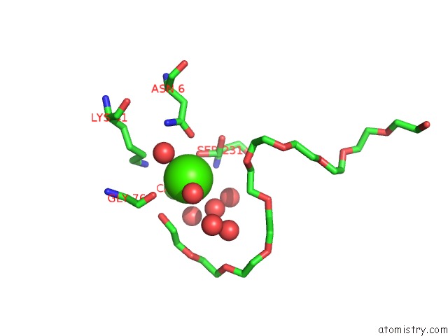
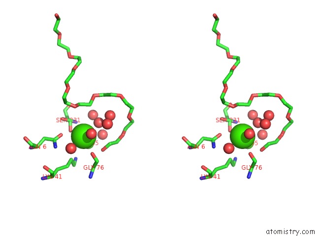
Calcium binding site 1 out of 6 in 7bip
Go back to
Calcium binding site 1 out
of 6 in the Crystal Structure of Monooxygenase RSLO1 From Streptomyces Bottropensis
Mono view
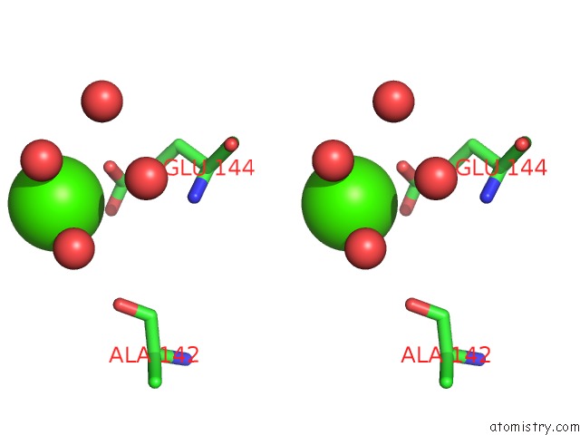
Stereo pair view
Mono view
Stereo pair view
A full contact list of Calcium with other atoms in the Ca binding
site number 1 of Crystal Structure of Monooxygenase RSLO1 From Streptomyces Bottropensis within 5.0Å range:
|
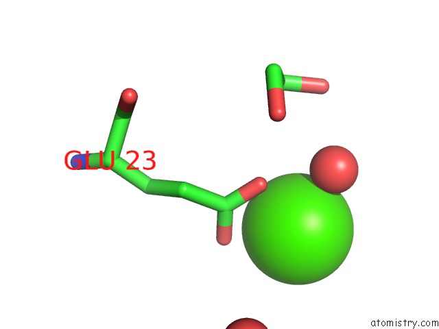
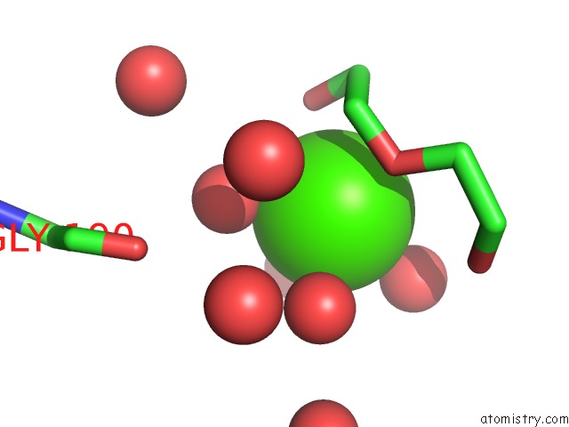
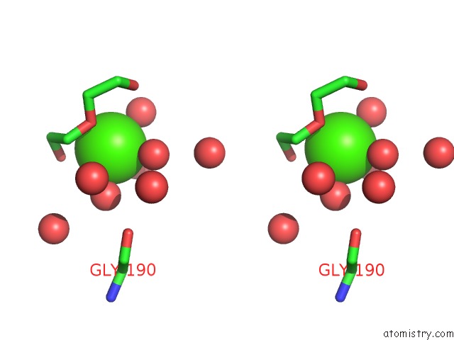
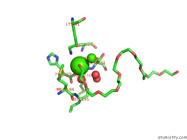
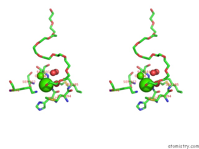
Calcium binding site 2 out of 6 in 7bip
Go back to
Calcium binding site 2 out
of 6 in the Crystal Structure of Monooxygenase RSLO1 From Streptomyces Bottropensis
Mono view
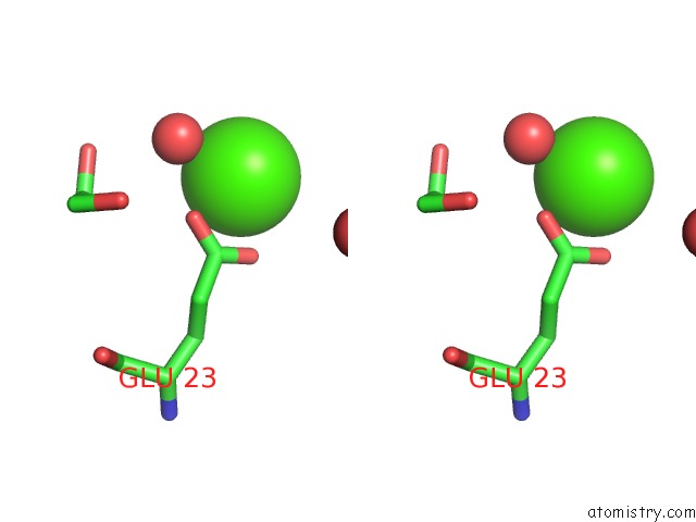
Stereo pair view
Mono view
Stereo pair view
A full contact list of Calcium with other atoms in the Ca binding
site number 2 of Crystal Structure of Monooxygenase RSLO1 From Streptomyces Bottropensis within 5.0Å range:
|
Calcium binding site 3 out of 6 in 7bip
Go back to
Calcium binding site 3 out
of 6 in the Crystal Structure of Monooxygenase RSLO1 From Streptomyces Bottropensis
Mono view
Stereo pair view
Mono view
Stereo pair view
A full contact list of Calcium with other atoms in the Ca binding
site number 3 of Crystal Structure of Monooxygenase RSLO1 From Streptomyces Bottropensis within 5.0Å range:
|
Calcium binding site 4 out of 6 in 7bip
Go back to
Calcium binding site 4 out
of 6 in the Crystal Structure of Monooxygenase RSLO1 From Streptomyces Bottropensis
Mono view
Stereo pair view
Mono view
Stereo pair view
A full contact list of Calcium with other atoms in the Ca binding
site number 4 of Crystal Structure of Monooxygenase RSLO1 From Streptomyces Bottropensis within 5.0Å range:
|
Calcium binding site 5 out of 6 in 7bip
Go back to
Calcium binding site 5 out
of 6 in the Crystal Structure of Monooxygenase RSLO1 From Streptomyces Bottropensis
Mono view
Stereo pair view
Mono view
Stereo pair view
A full contact list of Calcium with other atoms in the Ca binding
site number 5 of Crystal Structure of Monooxygenase RSLO1 From Streptomyces Bottropensis within 5.0Å range:
|
Calcium binding site 6 out of 6 in 7bip
Go back to
Calcium binding site 6 out
of 6 in the Crystal Structure of Monooxygenase RSLO1 From Streptomyces Bottropensis
Mono view
Stereo pair view
Mono view
Stereo pair view
A full contact list of Calcium with other atoms in the Ca binding
site number 6 of Crystal Structure of Monooxygenase RSLO1 From Streptomyces Bottropensis within 5.0Å range:
|
Reference:
A.Alali,
L.Zhang,
J.Li,
C.Zuo,
D.Wassouf,
X.Yan,
P.Schwarzer,
S.Guenther,
O.Einsle,
A.Bechthold.
Unique Third Ring Oxygenation After Three Cyclization Steps in the Biosynthesis of the Tricyclic Aromatic Pks-II Rishirilides To Be Published.
Page generated: Thu Jul 18 23:17:53 2024
Last articles
Zn in 9MJ5Zn in 9HNW
Zn in 9G0L
Zn in 9FNE
Zn in 9DZN
Zn in 9E0I
Zn in 9D32
Zn in 9DAK
Zn in 8ZXC
Zn in 8ZUF