Calcium »
PDB 7d52-7dl7 »
7d7e »
Calcium in PDB 7d7e: Structure of PKD1L3-Ctd/PKD2L1 in Apo State
Calcium Binding Sites:
The binding sites of Calcium atom in the Structure of PKD1L3-Ctd/PKD2L1 in Apo State
(pdb code 7d7e). This binding sites where shown within
5.0 Angstroms radius around Calcium atom.
In total 3 binding sites of Calcium where determined in the Structure of PKD1L3-Ctd/PKD2L1 in Apo State, PDB code: 7d7e:
Jump to Calcium binding site number: 1; 2; 3;
In total 3 binding sites of Calcium where determined in the Structure of PKD1L3-Ctd/PKD2L1 in Apo State, PDB code: 7d7e:
Jump to Calcium binding site number: 1; 2; 3;
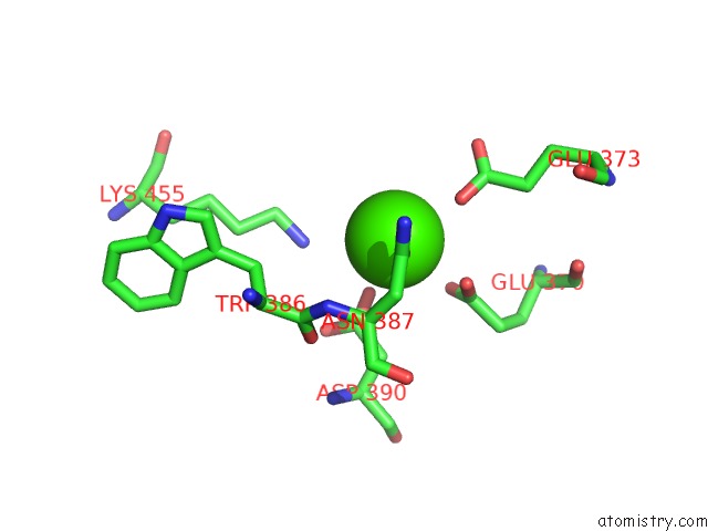
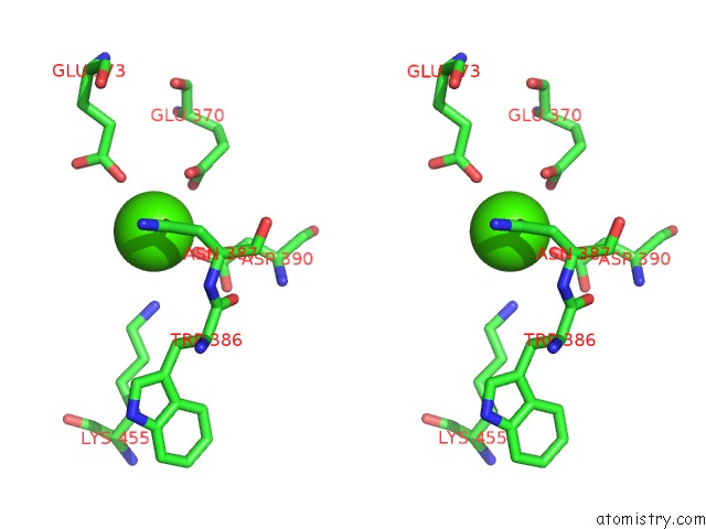
Calcium binding site 1 out of 3 in 7d7e
Go back to
Calcium binding site 1 out
of 3 in the Structure of PKD1L3-Ctd/PKD2L1 in Apo State
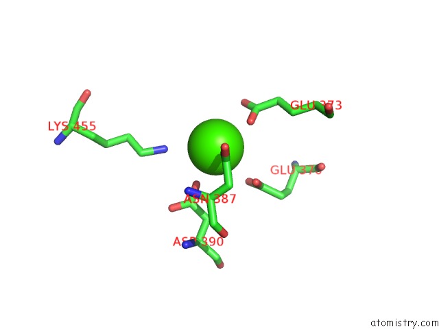
Mono view
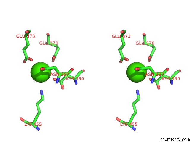
Stereo pair view
Mono view
Stereo pair view
A full contact list of Calcium with other atoms in the Ca binding
site number 1 of Structure of PKD1L3-Ctd/PKD2L1 in Apo State within 5.0Å range:
|
Calcium binding site 2 out of 3 in 7d7e
Go back to
Calcium binding site 2 out
of 3 in the Structure of PKD1L3-Ctd/PKD2L1 in Apo State
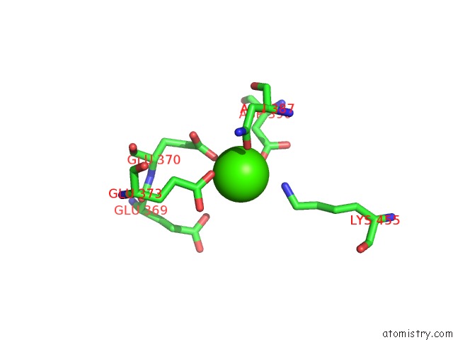
Mono view
Stereo pair view
Mono view
Stereo pair view
A full contact list of Calcium with other atoms in the Ca binding
site number 2 of Structure of PKD1L3-Ctd/PKD2L1 in Apo State within 5.0Å range:
|
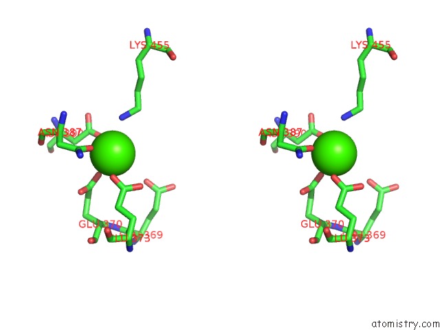
Calcium binding site 3 out of 3 in 7d7e
Go back to
Calcium binding site 3 out
of 3 in the Structure of PKD1L3-Ctd/PKD2L1 in Apo State
Mono view
Stereo pair view
Mono view
Stereo pair view
A full contact list of Calcium with other atoms in the Ca binding
site number 3 of Structure of PKD1L3-Ctd/PKD2L1 in Apo State within 5.0Å range:
|
Reference:
Q.Su,
M.Chen,
Y.Wang,
B.Li,
D.Jing,
X.Zhan,
Y.Yu,
Y.Shi.
Structural Basis For Ca 2+ Activation of the Heteromeric PKD1L3/PKD2L1 Channel. Nat Commun V. 12 4871 2021.
ISSN: ESSN 2041-1723
PubMed: 34381056
DOI: 10.1038/S41467-021-25216-Z
Page generated: Wed Jul 9 21:29:13 2025
ISSN: ESSN 2041-1723
PubMed: 34381056
DOI: 10.1038/S41467-021-25216-Z
Last articles
Cd in 2BT7Cd in 2BN0
Cd in 2BJD
Cd in 2B3P
Cd in 2BMZ
Cd in 2BMY
Cd in 2A97
Cd in 1ZWW
Cd in 2AVP
Cd in 2A38