Calcium »
PDB 7d4p-7dkx »
7dae »
Calcium in PDB 7dae: Epb in Complex with Tubulin
Protein crystallography data
The structure of Epb in Complex with Tubulin, PDB code: 7dae
was solved by
C.Wu,
Y.Wang,
with X-Ray Crystallography technique. A brief refinement statistics is given in the table below:
| Resolution Low / High (Å) | 49.71 / 2.39 |
| Space group | P 21 21 21 |
| Cell size a, b, c (Å), α, β, γ (°) | 104.792, 157.21, 181.53, 90, 90, 90 |
| R / Rfree (%) | 17.4 / 22.2 |
Other elements in 7dae:
The structure of Epb in Complex with Tubulin also contains other interesting chemical elements:
| Magnesium | (Mg) | 4 atoms |
| Chlorine | (Cl) | 1 atom |
Calcium Binding Sites:
The binding sites of Calcium atom in the Epb in Complex with Tubulin
(pdb code 7dae). This binding sites where shown within
5.0 Angstroms radius around Calcium atom.
In total 4 binding sites of Calcium where determined in the Epb in Complex with Tubulin, PDB code: 7dae:
Jump to Calcium binding site number: 1; 2; 3; 4;
In total 4 binding sites of Calcium where determined in the Epb in Complex with Tubulin, PDB code: 7dae:
Jump to Calcium binding site number: 1; 2; 3; 4;
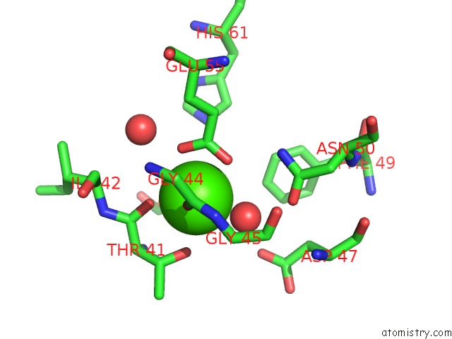
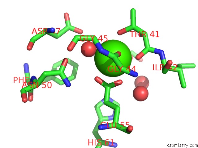
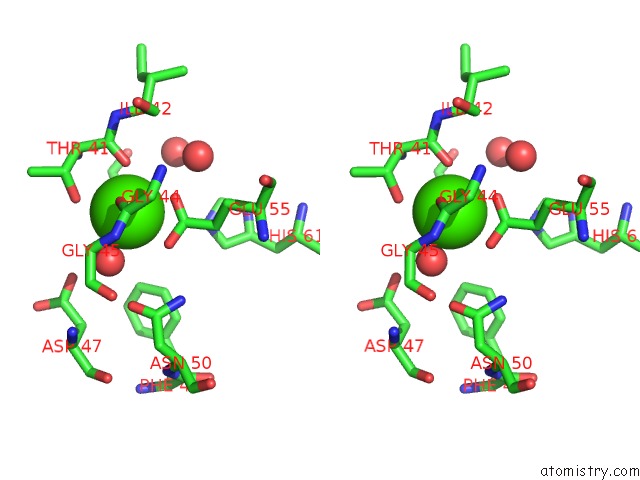
Calcium binding site 1 out of 4 in 7dae
Go back to
Calcium binding site 1 out
of 4 in the Epb in Complex with Tubulin
Mono view
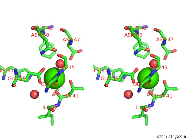
Stereo pair view
Mono view
Stereo pair view
A full contact list of Calcium with other atoms in the Ca binding
site number 1 of Epb in Complex with Tubulin within 5.0Å range:
|
Calcium binding site 2 out of 4 in 7dae
Go back to
Calcium binding site 2 out
of 4 in the Epb in Complex with Tubulin
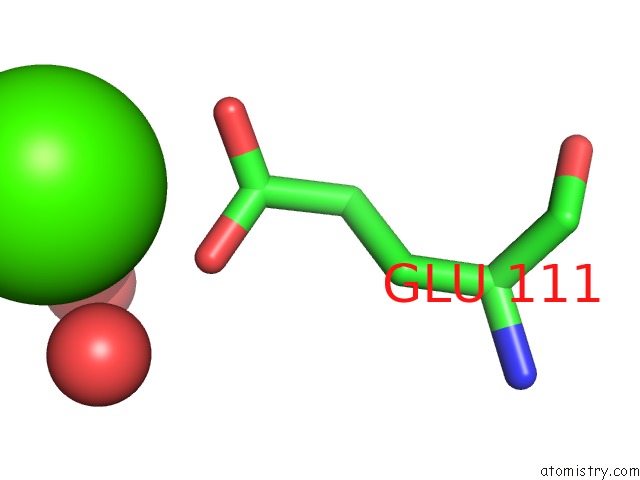
Mono view
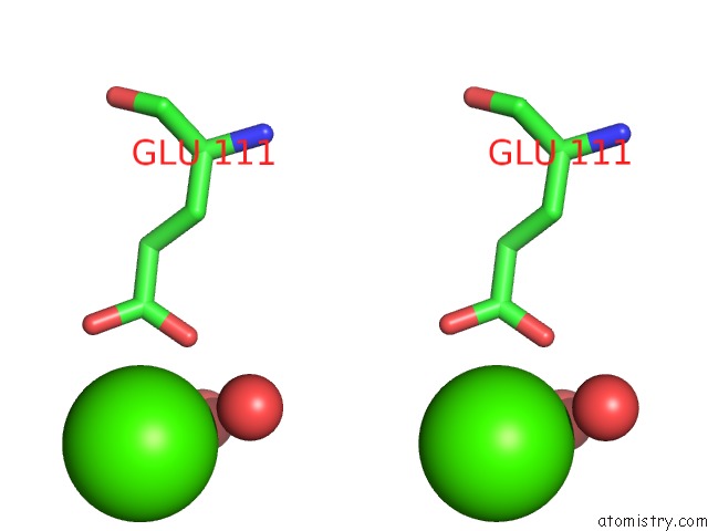
Stereo pair view
Mono view
Stereo pair view
A full contact list of Calcium with other atoms in the Ca binding
site number 2 of Epb in Complex with Tubulin within 5.0Å range:
|
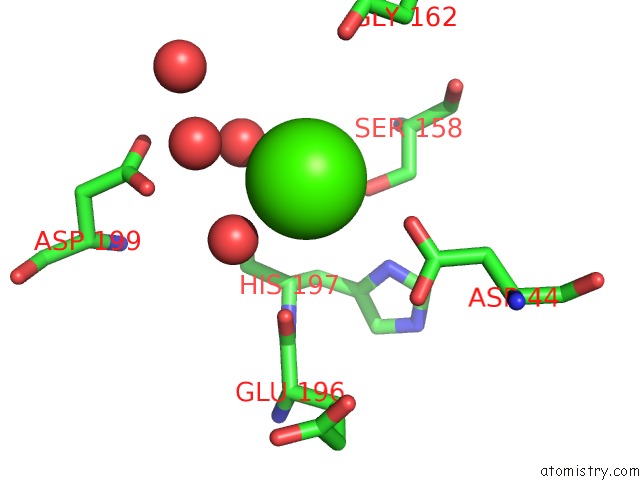
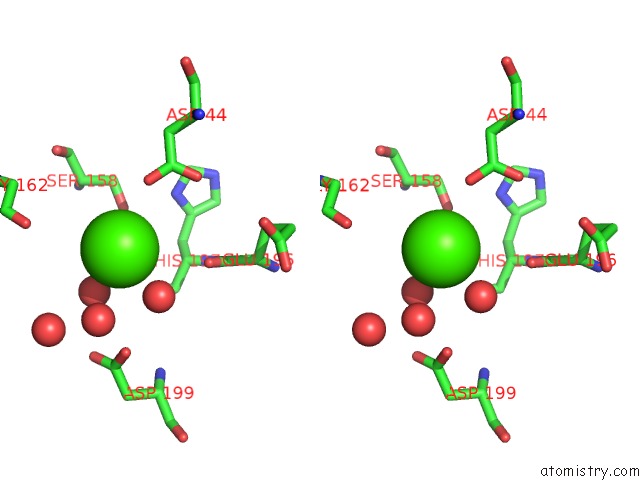
Calcium binding site 3 out of 4 in 7dae
Go back to
Calcium binding site 3 out
of 4 in the Epb in Complex with Tubulin
Mono view
Stereo pair view
Mono view
Stereo pair view
A full contact list of Calcium with other atoms in the Ca binding
site number 3 of Epb in Complex with Tubulin within 5.0Å range:
|
Calcium binding site 4 out of 4 in 7dae
Go back to
Calcium binding site 4 out
of 4 in the Epb in Complex with Tubulin
Mono view
Stereo pair view
Mono view
Stereo pair view
A full contact list of Calcium with other atoms in the Ca binding
site number 4 of Epb in Complex with Tubulin within 5.0Å range:
|
Reference:
Q.Xiao,
T.Xue,
W.Shuai,
C.Wu,
Z.Zhang,
T.Zhang,
S.Zeng,
B.Sun,
Y.Wang.
High-Resolution X-Ray Structure of Three Microtubule-Stabilizing Agents in Complex with Tubulin Provide A Rationale For Drug Design. Biochem.Biophys.Res.Commun. V. 534 330 2021.
ISSN: ESSN 1090-2104
PubMed: 33272565
DOI: 10.1016/J.BBRC.2020.11.082
Page generated: Thu Jul 18 23:59:48 2024
ISSN: ESSN 1090-2104
PubMed: 33272565
DOI: 10.1016/J.BBRC.2020.11.082
Last articles
Zn in 9JYWZn in 9IR4
Zn in 9IR3
Zn in 9GMX
Zn in 9GMW
Zn in 9JEJ
Zn in 9ERF
Zn in 9ERE
Zn in 9EGV
Zn in 9EGW