Calcium »
PDB 7dlh-7e6q »
7dmr »
Calcium in PDB 7dmr: Crystal Structure of Potassium Induced Heme Modification in Yak Lactoperoxidase at 2.20 A Resolution
Enzymatic activity of Crystal Structure of Potassium Induced Heme Modification in Yak Lactoperoxidase at 2.20 A Resolution
All present enzymatic activity of Crystal Structure of Potassium Induced Heme Modification in Yak Lactoperoxidase at 2.20 A Resolution:
1.11.1.7;
1.11.1.7;
Protein crystallography data
The structure of Crystal Structure of Potassium Induced Heme Modification in Yak Lactoperoxidase at 2.20 A Resolution, PDB code: 7dmr
was solved by
P.K.Singh,
C.Rani,
P.Sharma,
S.Sharma,
T.P.Singh,
with X-Ray Crystallography technique. A brief refinement statistics is given in the table below:
| Resolution Low / High (Å) | 64.61 / 2.20 |
| Space group | P 21 21 21 |
| Cell size a, b, c (Å), α, β, γ (°) | 80.240, 85.310, 98.930, 90.00, 90.00, 90.00 |
| R / Rfree (%) | 19 / 24.3 |
Other elements in 7dmr:
The structure of Crystal Structure of Potassium Induced Heme Modification in Yak Lactoperoxidase at 2.20 A Resolution also contains other interesting chemical elements:
| Potassium | (K) | 1 atom |
| Zinc | (Zn) | 2 atoms |
| Iron | (Fe) | 1 atom |
| Chlorine | (Cl) | 1 atom |
Calcium Binding Sites:
The binding sites of Calcium atom in the Crystal Structure of Potassium Induced Heme Modification in Yak Lactoperoxidase at 2.20 A Resolution
(pdb code 7dmr). This binding sites where shown within
5.0 Angstroms radius around Calcium atom.
In total only one binding site of Calcium was determined in the Crystal Structure of Potassium Induced Heme Modification in Yak Lactoperoxidase at 2.20 A Resolution, PDB code: 7dmr:
In total only one binding site of Calcium was determined in the Crystal Structure of Potassium Induced Heme Modification in Yak Lactoperoxidase at 2.20 A Resolution, PDB code: 7dmr:
Calcium binding site 1 out of 1 in 7dmr
Go back to
Calcium binding site 1 out
of 1 in the Crystal Structure of Potassium Induced Heme Modification in Yak Lactoperoxidase at 2.20 A Resolution
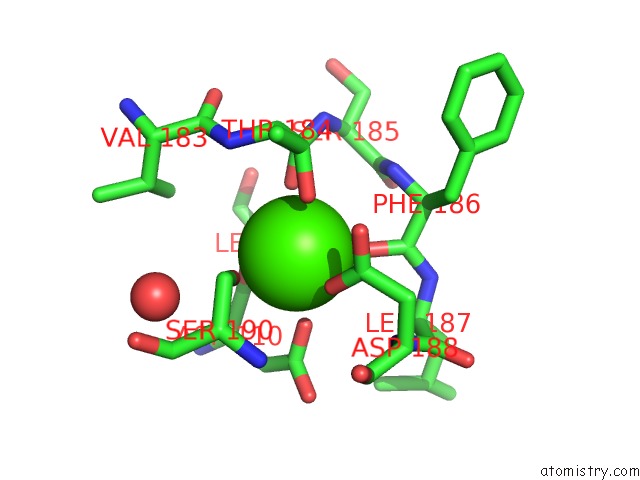
Mono view
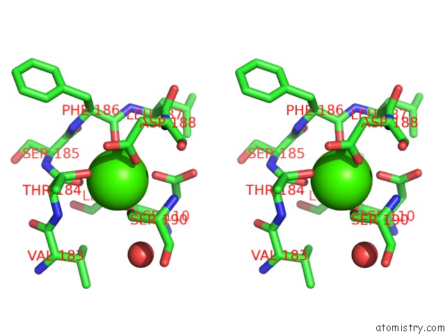
Stereo pair view
Mono view
Stereo pair view
A full contact list of Calcium with other atoms in the Ca binding
site number 1 of Crystal Structure of Potassium Induced Heme Modification in Yak Lactoperoxidase at 2.20 A Resolution within 5.0Å range:
|
Reference:
P.K.Singh,
C.Rani,
P.Sharma,
S.Shrama,
T.P.Singh.
Crystal Structure of Potassium Induced Heme Modification in Yak Lactoperoxidase at 2.20 A Resolution To Be Published.
Page generated: Wed Jul 9 21:38:19 2025
Last articles
Ca in 7G5SCa in 7G5Q
Ca in 7G5N
Ca in 7G5P
Ca in 7G5O
Ca in 7G5M
Ca in 7G5L
Ca in 7G5K
Ca in 7G5J
Ca in 7G5I