Calcium »
PDB 7dl7-7e4z »
7do4 »
Calcium in PDB 7do4: Crystal Structure of CD97-CD55 Complex
Protein crystallography data
The structure of Crystal Structure of CD97-CD55 Complex, PDB code: 7do4
was solved by
M.Niu,
G.Song,
with X-Ray Crystallography technique. A brief refinement statistics is given in the table below:
| Resolution Low / High (Å) | 33.23 / 3.20 |
| Space group | P 1 21 1 |
| Cell size a, b, c (Å), α, β, γ (°) | 51.8, 44.25, 116.53, 90, 98.7, 90 |
| R / Rfree (%) | 26.7 / 30.4 |
Calcium Binding Sites:
The binding sites of Calcium atom in the Crystal Structure of CD97-CD55 Complex
(pdb code 7do4). This binding sites where shown within
5.0 Angstroms radius around Calcium atom.
In total 2 binding sites of Calcium where determined in the Crystal Structure of CD97-CD55 Complex, PDB code: 7do4:
Jump to Calcium binding site number: 1; 2;
In total 2 binding sites of Calcium where determined in the Crystal Structure of CD97-CD55 Complex, PDB code: 7do4:
Jump to Calcium binding site number: 1; 2;
Calcium binding site 1 out of 2 in 7do4
Go back to
Calcium binding site 1 out
of 2 in the Crystal Structure of CD97-CD55 Complex
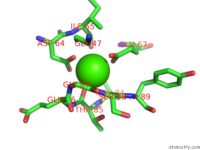
Mono view
Stereo pair view
Mono view
Stereo pair view
A full contact list of Calcium with other atoms in the Ca binding
site number 1 of Crystal Structure of CD97-CD55 Complex within 5.0Å range:
|
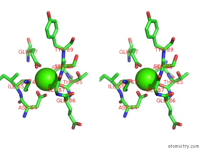
Calcium binding site 2 out of 2 in 7do4
Go back to
Calcium binding site 2 out
of 2 in the Crystal Structure of CD97-CD55 Complex

Mono view

Stereo pair view
Mono view
Stereo pair view
A full contact list of Calcium with other atoms in the Ca binding
site number 2 of Crystal Structure of CD97-CD55 Complex within 5.0Å range:
|
Reference:
M.Niu,
S.Xu,
J.Yang,
D.Yao,
N.Li,
J.Yan,
G.Zhong,
G.Song.
Structural Basis For CD97 Recognition of the Decay-Accelerating Factor CD55 Suggests Mechanosensitive Activation of Adhesion Gpcrs. J.Biol.Chem. V. 296 00776 2021.
ISSN: ESSN 1083-351X
PubMed: 33992645
DOI: 10.1016/J.JBC.2021.100776
Page generated: Fri Jul 19 00:08:22 2024
ISSN: ESSN 1083-351X
PubMed: 33992645
DOI: 10.1016/J.JBC.2021.100776
Last articles
Zn in 9J0NZn in 9J0O
Zn in 9J0P
Zn in 9FJX
Zn in 9EKB
Zn in 9C0F
Zn in 9CAH
Zn in 9CH0
Zn in 9CH3
Zn in 9CH1