Calcium »
PDB 7dl7-7e4z »
7dq6 »
Calcium in PDB 7dq6: Crystal Structure of Hitb in Complex with (S)-Beta-3-Br-Phenylalanine Sulfamoyladenosine
Protein crystallography data
The structure of Crystal Structure of Hitb in Complex with (S)-Beta-3-Br-Phenylalanine Sulfamoyladenosine, PDB code: 7dq6
was solved by
F.Kudo,
S.Takahashi,
A.Miyanaga,
Y.Nakazawa,
T.Eguchi,
with X-Ray Crystallography technique. A brief refinement statistics is given in the table below:
| Resolution Low / High (Å) | 48.39 / 2.60 |
| Space group | P 21 21 21 |
| Cell size a, b, c (Å), α, β, γ (°) | 67.965, 96.693, 166.414, 90, 90, 90 |
| R / Rfree (%) | 21.2 / 27 |
Other elements in 7dq6:
The structure of Crystal Structure of Hitb in Complex with (S)-Beta-3-Br-Phenylalanine Sulfamoyladenosine also contains other interesting chemical elements:
| Bromine | (Br) | 2 atoms |
Calcium Binding Sites:
The binding sites of Calcium atom in the Crystal Structure of Hitb in Complex with (S)-Beta-3-Br-Phenylalanine Sulfamoyladenosine
(pdb code 7dq6). This binding sites where shown within
5.0 Angstroms radius around Calcium atom.
In total 2 binding sites of Calcium where determined in the Crystal Structure of Hitb in Complex with (S)-Beta-3-Br-Phenylalanine Sulfamoyladenosine, PDB code: 7dq6:
Jump to Calcium binding site number: 1; 2;
In total 2 binding sites of Calcium where determined in the Crystal Structure of Hitb in Complex with (S)-Beta-3-Br-Phenylalanine Sulfamoyladenosine, PDB code: 7dq6:
Jump to Calcium binding site number: 1; 2;
Calcium binding site 1 out of 2 in 7dq6
Go back to
Calcium binding site 1 out
of 2 in the Crystal Structure of Hitb in Complex with (S)-Beta-3-Br-Phenylalanine Sulfamoyladenosine
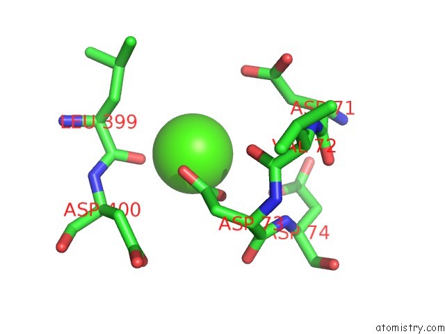
Mono view
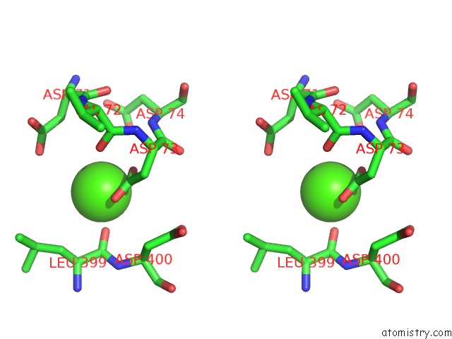
Stereo pair view
Mono view
Stereo pair view
A full contact list of Calcium with other atoms in the Ca binding
site number 1 of Crystal Structure of Hitb in Complex with (S)-Beta-3-Br-Phenylalanine Sulfamoyladenosine within 5.0Å range:
|
Calcium binding site 2 out of 2 in 7dq6
Go back to
Calcium binding site 2 out
of 2 in the Crystal Structure of Hitb in Complex with (S)-Beta-3-Br-Phenylalanine Sulfamoyladenosine
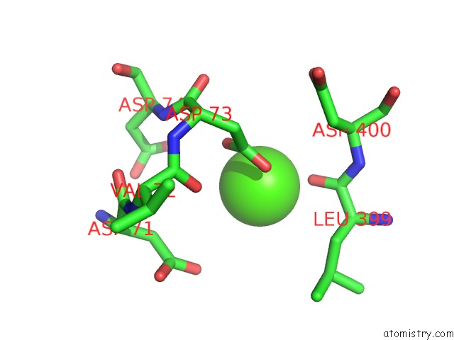
Mono view
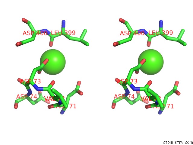
Stereo pair view
Mono view
Stereo pair view
A full contact list of Calcium with other atoms in the Ca binding
site number 2 of Crystal Structure of Hitb in Complex with (S)-Beta-3-Br-Phenylalanine Sulfamoyladenosine within 5.0Å range:
|
Reference:
F.Kudo,
S.Takahashi,
A.Miyanaga,
Y.Nakazawa,
K.Nishino,
Y.Hayakawa,
K.Kawamura,
F.Ishikawa,
G.Tanabe,
N.Iwai,
Y.Nagumo,
T.Usui,
T.Eguchi.
Mutational Biosynthesis of Hitachimycin Analogs Controlled By the Beta-Amino Acid-Selective Adenylation Enzyme Hitb. Acs Chem.Biol. 2021.
ISSN: ESSN 1554-8937
PubMed: 33625847
DOI: 10.1021/ACSCHEMBIO.1C00003
Page generated: Fri Jul 19 00:09:15 2024
ISSN: ESSN 1554-8937
PubMed: 33625847
DOI: 10.1021/ACSCHEMBIO.1C00003
Last articles
Zn in 9J0NZn in 9J0O
Zn in 9J0P
Zn in 9FJX
Zn in 9EKB
Zn in 9C0F
Zn in 9CAH
Zn in 9CH0
Zn in 9CH3
Zn in 9CH1