Calcium »
PDB 7dlh-7e6q »
7dt8 »
Calcium in PDB 7dt8: Crystal Structure of V30M Mutated Transthyretin in Complex with 4- Chloro-9-Oxo-9H-Xanthene-2-Carboxylic Acid
Protein crystallography data
The structure of Crystal Structure of V30M Mutated Transthyretin in Complex with 4- Chloro-9-Oxo-9H-Xanthene-2-Carboxylic Acid, PDB code: 7dt8
was solved by
R.Kitakami,
T.Yokoyama,
M.Mizuguchi,
with X-Ray Crystallography technique. A brief refinement statistics is given in the table below:
| Resolution Low / High (Å) | 35.50 / 1.25 |
| Space group | P 21 21 2 |
| Cell size a, b, c (Å), α, β, γ (°) | 42.942, 85.839, 63.282, 90, 90, 90 |
| R / Rfree (%) | 16.1 / 17.7 |
Other elements in 7dt8:
The structure of Crystal Structure of V30M Mutated Transthyretin in Complex with 4- Chloro-9-Oxo-9H-Xanthene-2-Carboxylic Acid also contains other interesting chemical elements:
| Chlorine | (Cl) | 2 atoms |
Calcium Binding Sites:
The binding sites of Calcium atom in the Crystal Structure of V30M Mutated Transthyretin in Complex with 4- Chloro-9-Oxo-9H-Xanthene-2-Carboxylic Acid
(pdb code 7dt8). This binding sites where shown within
5.0 Angstroms radius around Calcium atom.
In total 2 binding sites of Calcium where determined in the Crystal Structure of V30M Mutated Transthyretin in Complex with 4- Chloro-9-Oxo-9H-Xanthene-2-Carboxylic Acid, PDB code: 7dt8:
Jump to Calcium binding site number: 1; 2;
In total 2 binding sites of Calcium where determined in the Crystal Structure of V30M Mutated Transthyretin in Complex with 4- Chloro-9-Oxo-9H-Xanthene-2-Carboxylic Acid, PDB code: 7dt8:
Jump to Calcium binding site number: 1; 2;
Calcium binding site 1 out of 2 in 7dt8
Go back to
Calcium binding site 1 out
of 2 in the Crystal Structure of V30M Mutated Transthyretin in Complex with 4- Chloro-9-Oxo-9H-Xanthene-2-Carboxylic Acid
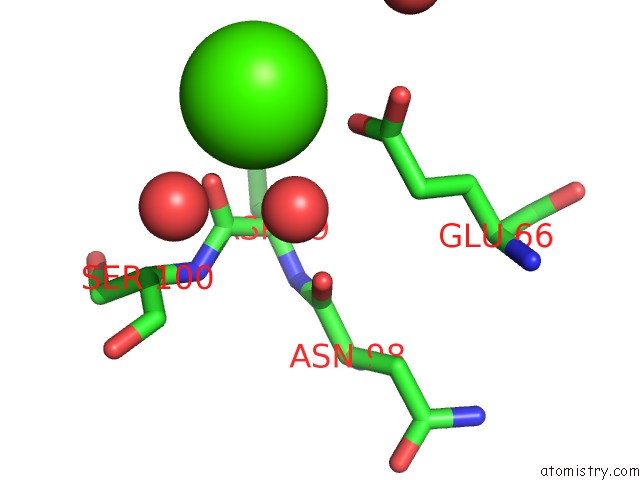
Mono view
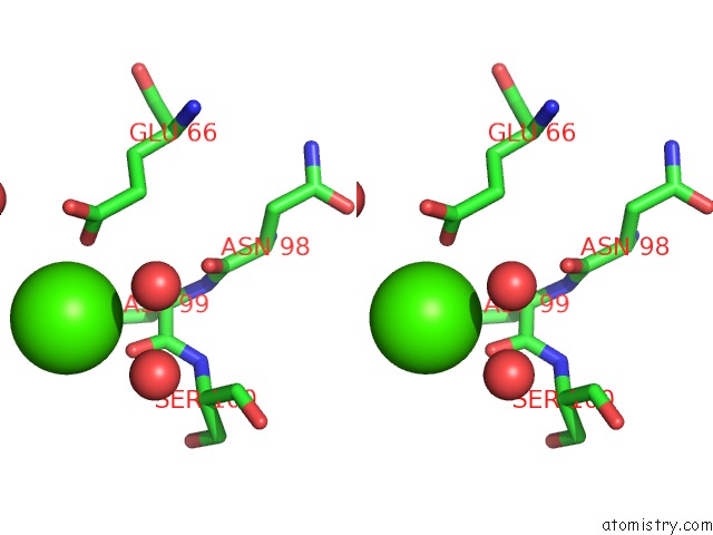
Stereo pair view
Mono view
Stereo pair view
A full contact list of Calcium with other atoms in the Ca binding
site number 1 of Crystal Structure of V30M Mutated Transthyretin in Complex with 4- Chloro-9-Oxo-9H-Xanthene-2-Carboxylic Acid within 5.0Å range:
|
Calcium binding site 2 out of 2 in 7dt8
Go back to
Calcium binding site 2 out
of 2 in the Crystal Structure of V30M Mutated Transthyretin in Complex with 4- Chloro-9-Oxo-9H-Xanthene-2-Carboxylic Acid
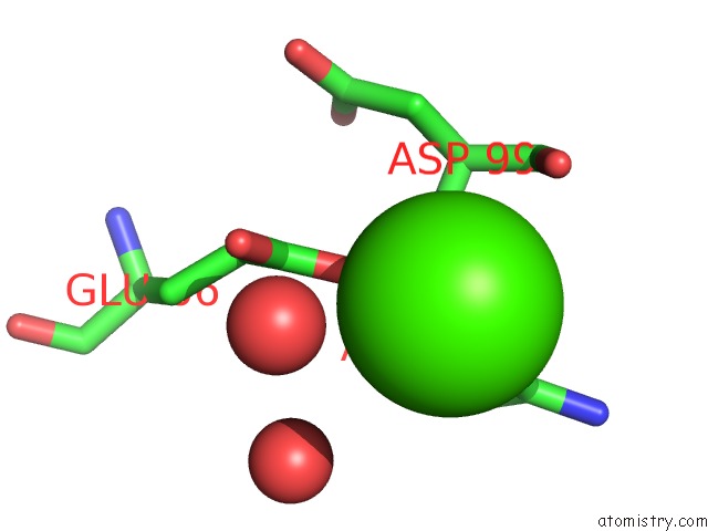
Mono view
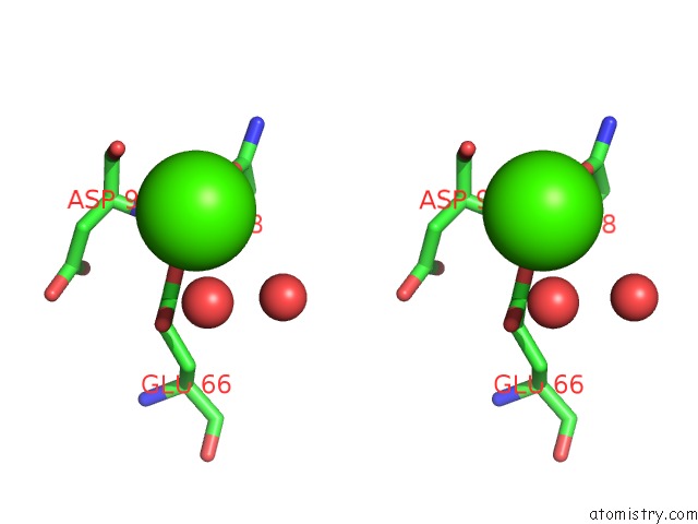
Stereo pair view
Mono view
Stereo pair view
A full contact list of Calcium with other atoms in the Ca binding
site number 2 of Crystal Structure of V30M Mutated Transthyretin in Complex with 4- Chloro-9-Oxo-9H-Xanthene-2-Carboxylic Acid within 5.0Å range:
|
Reference:
R.Kitakami,
K.Inui,
Y.Nakagawa,
Y.Sawai,
W.Katayama,
T.Yokoyama,
T.Okada,
K.Kanamitsu,
S.Nakagawa,
N.Toyooka,
M.Mizuguchi.
Inhibitory Activities of Anthraquinone and Xanthone Derivatives Against Transthyretin Amyloidogenesis. Bioorg.Med.Chem. V. 44 16292 2021.
ISSN: ESSN 1464-3391
PubMed: 34225167
DOI: 10.1016/J.BMC.2021.116292
Page generated: Wed Jul 9 21:40:28 2025
ISSN: ESSN 1464-3391
PubMed: 34225167
DOI: 10.1016/J.BMC.2021.116292
Last articles
Fe in 2YXOFe in 2YRS
Fe in 2YXC
Fe in 2YNM
Fe in 2YVJ
Fe in 2YP1
Fe in 2YU2
Fe in 2YU1
Fe in 2YQB
Fe in 2YOO