Calcium »
PDB 7e6q-7esy »
7e9y »
Calcium in PDB 7e9y: Crystal Structure of ELACCO1
Protein crystallography data
The structure of Crystal Structure of ELACCO1, PDB code: 7e9y
was solved by
Y.Wen,
R.E.Campbell,
M.J.Lemieux,
Y.Nasu,
with X-Ray Crystallography technique. A brief refinement statistics is given in the table below:
| Resolution Low / High (Å) | 52.49 / 2.25 |
| Space group | C 2 2 21 |
| Cell size a, b, c (Å), α, β, γ (°) | 102.217, 104.983, 138.748, 90, 90, 90 |
| R / Rfree (%) | 14.7 / 18.5 |
Calcium Binding Sites:
The binding sites of Calcium atom in the Crystal Structure of ELACCO1
(pdb code 7e9y). This binding sites where shown within
5.0 Angstroms radius around Calcium atom.
In total only one binding site of Calcium was determined in the Crystal Structure of ELACCO1, PDB code: 7e9y:
In total only one binding site of Calcium was determined in the Crystal Structure of ELACCO1, PDB code: 7e9y:
Calcium binding site 1 out of 1 in 7e9y
Go back to
Calcium binding site 1 out
of 1 in the Crystal Structure of ELACCO1
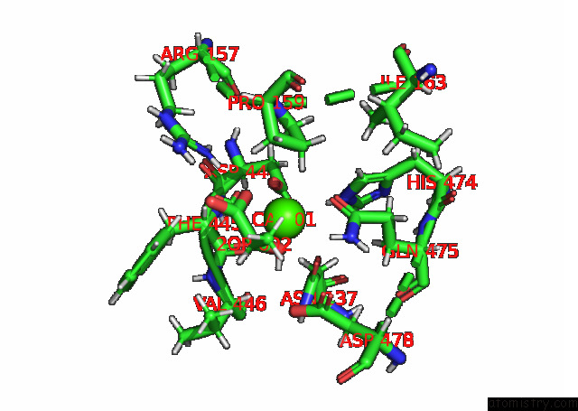
Mono view
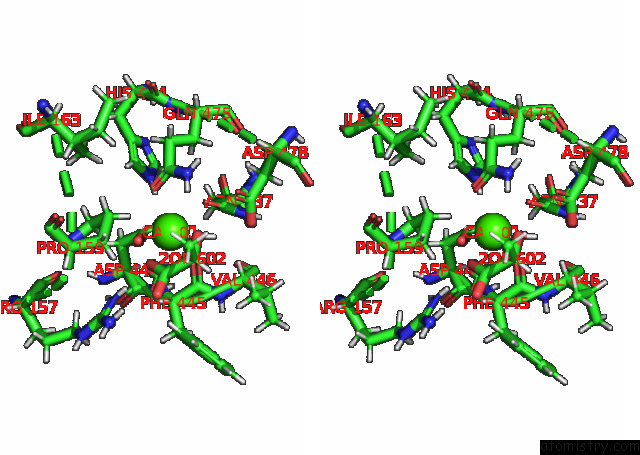
Stereo pair view
Mono view
Stereo pair view
A full contact list of Calcium with other atoms in the Ca binding
site number 1 of Crystal Structure of ELACCO1 within 5.0Å range:
|
Reference:
Y.Nasu,
C.Murphy-Royal,
Y.Wen,
J.N.Haidey,
R.S.Molina,
A.Aggarwal,
S.Zhang,
Y.Kamijo,
M.E.Paquet,
K.Podgorski,
M.Drobizhev,
J.S.Bains,
M.J.Lemieux,
G.R.Gordon,
R.E.Campbell.
A Genetically Encoded Fluorescent Biosensor For Extracellular L-Lactate. Nat Commun V. 12 7058 2021.
ISSN: ESSN 2041-1723
PubMed: 34873165
DOI: 10.1038/S41467-021-27332-2
Page generated: Fri Jul 19 00:20:03 2024
ISSN: ESSN 2041-1723
PubMed: 34873165
DOI: 10.1038/S41467-021-27332-2
Last articles
Zn in 9J0NZn in 9J0O
Zn in 9J0P
Zn in 9FJX
Zn in 9EKB
Zn in 9C0F
Zn in 9CAH
Zn in 9CH0
Zn in 9CH3
Zn in 9CH1