Calcium »
PDB 7e6q-7esy »
7ea4 »
Calcium in PDB 7ea4: Crystal Structure of L182E D-Succinylase (Dsa) From Cupriavidus Sp. P4-10-C
Protein crystallography data
The structure of Crystal Structure of L182E D-Succinylase (Dsa) From Cupriavidus Sp. P4-10-C, PDB code: 7ea4
was solved by
M.Yamasaki,
Y.Sumida,
with X-Ray Crystallography technique. A brief refinement statistics is given in the table below:
| Resolution Low / High (Å) | 42.93 / 1.95 |
| Space group | P 21 21 21 |
| Cell size a, b, c (Å), α, β, γ (°) | 62.057, 129.806, 200.68, 90, 90, 90 |
| R / Rfree (%) | 16.4 / 20.8 |
Other elements in 7ea4:
The structure of Crystal Structure of L182E D-Succinylase (Dsa) From Cupriavidus Sp. P4-10-C also contains other interesting chemical elements:
| Chlorine | (Cl) | 2 atoms |
| Arsenic | (As) | 2 atoms |
Calcium Binding Sites:
The binding sites of Calcium atom in the Crystal Structure of L182E D-Succinylase (Dsa) From Cupriavidus Sp. P4-10-C
(pdb code 7ea4). This binding sites where shown within
5.0 Angstroms radius around Calcium atom.
In total 2 binding sites of Calcium where determined in the Crystal Structure of L182E D-Succinylase (Dsa) From Cupriavidus Sp. P4-10-C, PDB code: 7ea4:
Jump to Calcium binding site number: 1; 2;
In total 2 binding sites of Calcium where determined in the Crystal Structure of L182E D-Succinylase (Dsa) From Cupriavidus Sp. P4-10-C, PDB code: 7ea4:
Jump to Calcium binding site number: 1; 2;
Calcium binding site 1 out of 2 in 7ea4
Go back to
Calcium binding site 1 out
of 2 in the Crystal Structure of L182E D-Succinylase (Dsa) From Cupriavidus Sp. P4-10-C
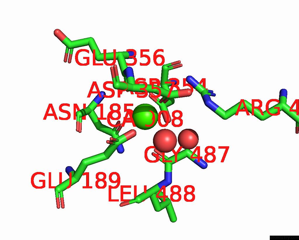
Mono view
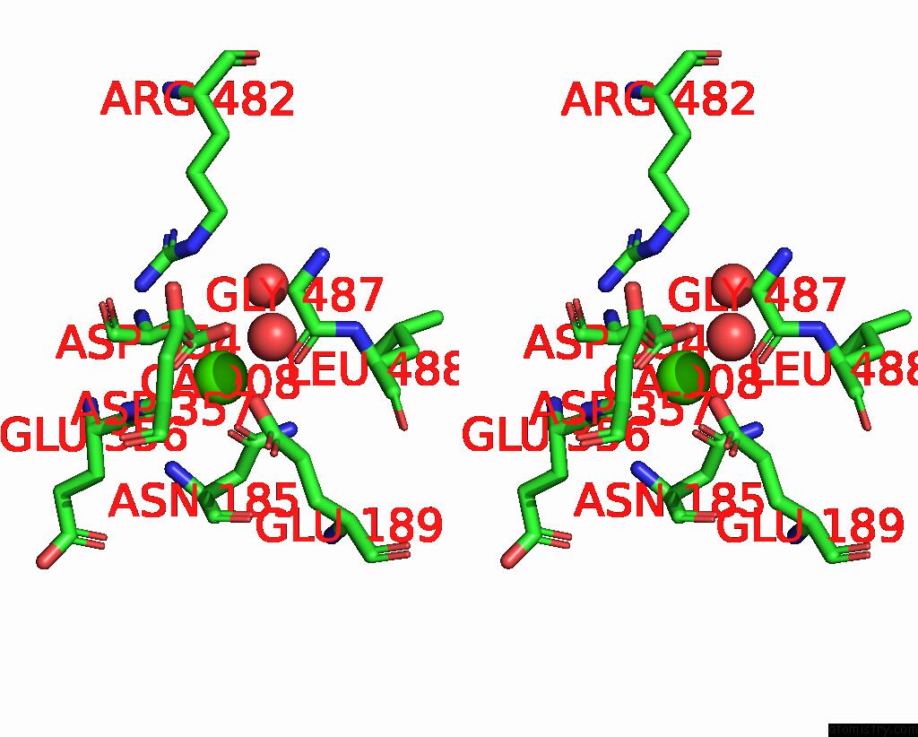
Stereo pair view
Mono view
Stereo pair view
A full contact list of Calcium with other atoms in the Ca binding
site number 1 of Crystal Structure of L182E D-Succinylase (Dsa) From Cupriavidus Sp. P4-10-C within 5.0Å range:
|
Calcium binding site 2 out of 2 in 7ea4
Go back to
Calcium binding site 2 out
of 2 in the Crystal Structure of L182E D-Succinylase (Dsa) From Cupriavidus Sp. P4-10-C
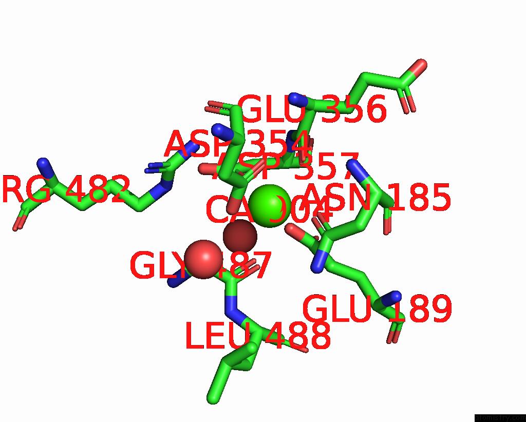
Mono view
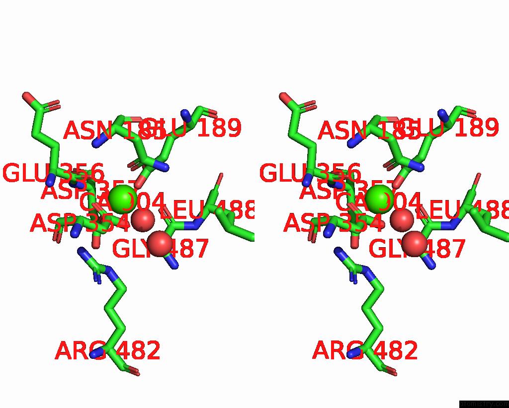
Stereo pair view
Mono view
Stereo pair view
A full contact list of Calcium with other atoms in the Ca binding
site number 2 of Crystal Structure of L182E D-Succinylase (Dsa) From Cupriavidus Sp. P4-10-C within 5.0Å range:
|
Reference:
Y.Sumida,
M.Yamasaki,
Y.Nishiya,
S.Kumagai,
T.Yamada,
M.Azuma.
Protein Engineering of D-Succinylase From Cupriavidus Sp. For D-Amino Acid Synthesis and the Structural Implications. Adv.Synth.Catal. V. 363 4770 2021.
ISSN: ESSN 1615-4169
DOI: 10.1002/ADSC.202100587
Page generated: Fri Jul 19 00:20:23 2024
ISSN: ESSN 1615-4169
DOI: 10.1002/ADSC.202100587
Last articles
Zn in 9J0NZn in 9J0O
Zn in 9J0P
Zn in 9FJX
Zn in 9EKB
Zn in 9C0F
Zn in 9CAH
Zn in 9CH0
Zn in 9CH3
Zn in 9CH1