Calcium »
PDB 7et8-7f9d »
7exo »
Calcium in PDB 7exo: Structure of Legume Lectin Domain From Methanocaldococcus Jannaschii in Mannose Bound Form
Protein crystallography data
The structure of Structure of Legume Lectin Domain From Methanocaldococcus Jannaschii in Mannose Bound Form, PDB code: 7exo
was solved by
K.Suguna,
F.Khan,
with X-Ray Crystallography technique. A brief refinement statistics is given in the table below:
| Resolution Low / High (Å) | 51.62 / 1.75 |
| Space group | P 41 21 2 |
| Cell size a, b, c (Å), α, β, γ (°) | 55.03, 55.03, 149.05, 90, 90, 90 |
| R / Rfree (%) | 15.9 / 18.3 |
Other elements in 7exo:
The structure of Structure of Legume Lectin Domain From Methanocaldococcus Jannaschii in Mannose Bound Form also contains other interesting chemical elements:
| Manganese | (Mn) | 1 atom |
Calcium Binding Sites:
The binding sites of Calcium atom in the Structure of Legume Lectin Domain From Methanocaldococcus Jannaschii in Mannose Bound Form
(pdb code 7exo). This binding sites where shown within
5.0 Angstroms radius around Calcium atom.
In total only one binding site of Calcium was determined in the Structure of Legume Lectin Domain From Methanocaldococcus Jannaschii in Mannose Bound Form, PDB code: 7exo:
In total only one binding site of Calcium was determined in the Structure of Legume Lectin Domain From Methanocaldococcus Jannaschii in Mannose Bound Form, PDB code: 7exo:
Calcium binding site 1 out of 1 in 7exo
Go back to
Calcium binding site 1 out
of 1 in the Structure of Legume Lectin Domain From Methanocaldococcus Jannaschii in Mannose Bound Form
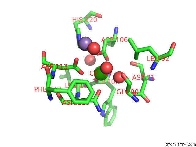
Mono view
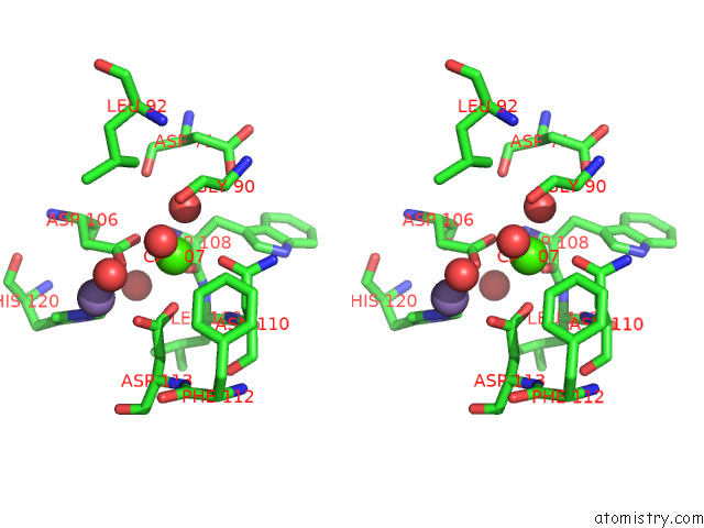
Stereo pair view
Mono view
Stereo pair view
A full contact list of Calcium with other atoms in the Ca binding
site number 1 of Structure of Legume Lectin Domain From Methanocaldococcus Jannaschii in Mannose Bound Form within 5.0Å range:
|
Reference:
F.Khan,
S.Kaza.
Crystal Structure of An L-Type Lectin Domain From Archaea. Proteins 2022.
ISSN: ESSN 1097-0134
PubMed: 36301308
DOI: 10.1002/PROT.26440
Page generated: Fri Jul 19 00:33:01 2024
ISSN: ESSN 1097-0134
PubMed: 36301308
DOI: 10.1002/PROT.26440
Last articles
Zn in 9J0NZn in 9J0O
Zn in 9J0P
Zn in 9FJX
Zn in 9EKB
Zn in 9C0F
Zn in 9CAH
Zn in 9CH0
Zn in 9CH3
Zn in 9CH1