Calcium »
PDB 7et9-7f9g »
7f4y »
Calcium in PDB 7f4y: Crystal Structure of Replisomal Dimer of Dna Polymerase From Bacteriophage RB69 with Dna Duplexes
Enzymatic activity of Crystal Structure of Replisomal Dimer of Dna Polymerase From Bacteriophage RB69 with Dna Duplexes
All present enzymatic activity of Crystal Structure of Replisomal Dimer of Dna Polymerase From Bacteriophage RB69 with Dna Duplexes:
2.7.7.7;
2.7.7.7;
Protein crystallography data
The structure of Crystal Structure of Replisomal Dimer of Dna Polymerase From Bacteriophage RB69 with Dna Duplexes, PDB code: 7f4y
was solved by
H.-S.Youn,
J.Park,
J.Y.An,
Y.Lee,
S.H.Eom,
J.Wang,
with X-Ray Crystallography technique. A brief refinement statistics is given in the table below:
| Resolution Low / High (Å) | 50.01 / 2.20 |
| Space group | P 32 2 1 |
| Cell size a, b, c (Å), α, β, γ (°) | 164.44, 164.44, 165.601, 90, 90, 120 |
| R / Rfree (%) | 19.4 / 24.5 |
Other elements in 7f4y:
The structure of Crystal Structure of Replisomal Dimer of Dna Polymerase From Bacteriophage RB69 with Dna Duplexes also contains other interesting chemical elements:
| Magnesium | (Mg) | 1 atom |
Calcium Binding Sites:
The binding sites of Calcium atom in the Crystal Structure of Replisomal Dimer of Dna Polymerase From Bacteriophage RB69 with Dna Duplexes
(pdb code 7f4y). This binding sites where shown within
5.0 Angstroms radius around Calcium atom.
In total 2 binding sites of Calcium where determined in the Crystal Structure of Replisomal Dimer of Dna Polymerase From Bacteriophage RB69 with Dna Duplexes, PDB code: 7f4y:
Jump to Calcium binding site number: 1; 2;
In total 2 binding sites of Calcium where determined in the Crystal Structure of Replisomal Dimer of Dna Polymerase From Bacteriophage RB69 with Dna Duplexes, PDB code: 7f4y:
Jump to Calcium binding site number: 1; 2;
Calcium binding site 1 out of 2 in 7f4y
Go back to
Calcium binding site 1 out
of 2 in the Crystal Structure of Replisomal Dimer of Dna Polymerase From Bacteriophage RB69 with Dna Duplexes
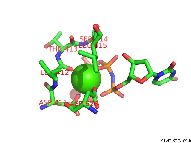
Mono view
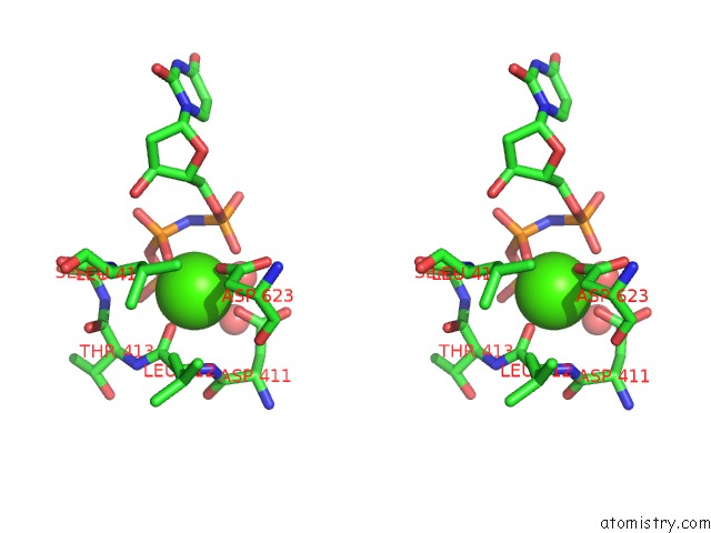
Stereo pair view
Mono view
Stereo pair view
A full contact list of Calcium with other atoms in the Ca binding
site number 1 of Crystal Structure of Replisomal Dimer of Dna Polymerase From Bacteriophage RB69 with Dna Duplexes within 5.0Å range:
|
Calcium binding site 2 out of 2 in 7f4y
Go back to
Calcium binding site 2 out
of 2 in the Crystal Structure of Replisomal Dimer of Dna Polymerase From Bacteriophage RB69 with Dna Duplexes
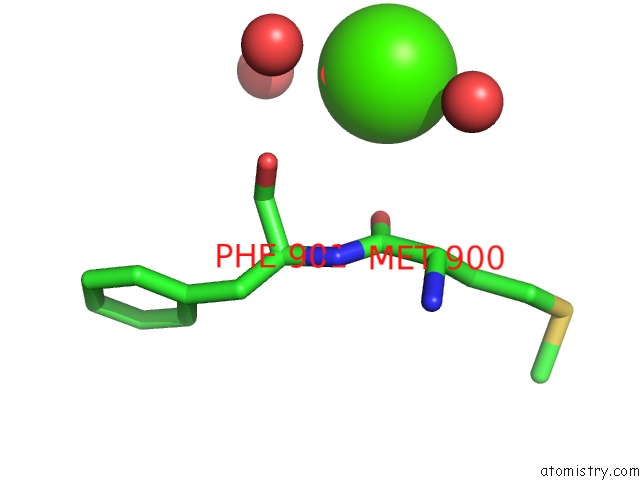
Mono view
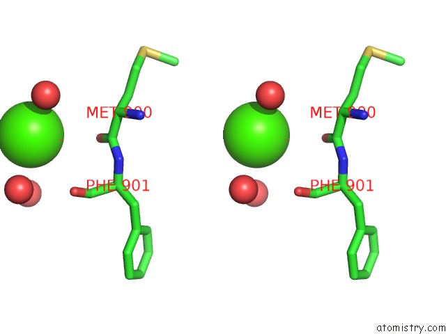
Stereo pair view
Mono view
Stereo pair view
A full contact list of Calcium with other atoms in the Ca binding
site number 2 of Crystal Structure of Replisomal Dimer of Dna Polymerase From Bacteriophage RB69 with Dna Duplexes within 5.0Å range:
|
Reference:
H.-S.Youn,
J.Park,
J.Y.An,
Y.Lee,
S.H.Eom,
J.Wang.
Crystal Structure of Replisomal Dimer of Dna Polymerase From Bacteriophage RB69 with Dna Duplexes To Be Published.
Page generated: Wed Jul 9 21:59:20 2025
Last articles
Cl in 5GRQCl in 5GRN
Cl in 5GSU
Cl in 5GS8
Cl in 5GQQ
Cl in 5G6U
Cl in 5GQN
Cl in 5GQM
Cl in 5GQL
Cl in 5GQK