Calcium »
PDB 7f9g-7g2q »
7fe1 »
Calcium in PDB 7fe1: Crystal Structure of GH92 Alpha-1,2-Mannosidase From Enterococcus Faecalis Atcc 10100 in Complex with Methyl Alpha-1,2-C-Mannobioside
Protein crystallography data
The structure of Crystal Structure of GH92 Alpha-1,2-Mannosidase From Enterococcus Faecalis Atcc 10100 in Complex with Methyl Alpha-1,2-C-Mannobioside, PDB code: 7fe1
was solved by
T.Miyazaki,
S.Alonso-Gil,
with X-Ray Crystallography technique. A brief refinement statistics is given in the table below:
| Resolution Low / High (Å) | 48.62 / 1.72 |
| Space group | C 2 2 21 |
| Cell size a, b, c (Å), α, β, γ (°) | 163.162, 169.601, 258.659, 90, 90, 90 |
| R / Rfree (%) | 14.9 / 17.1 |
Other elements in 7fe1:
The structure of Crystal Structure of GH92 Alpha-1,2-Mannosidase From Enterococcus Faecalis Atcc 10100 in Complex with Methyl Alpha-1,2-C-Mannobioside also contains other interesting chemical elements:
| Sodium | (Na) | 4 atoms |
Calcium Binding Sites:
The binding sites of Calcium atom in the Crystal Structure of GH92 Alpha-1,2-Mannosidase From Enterococcus Faecalis Atcc 10100 in Complex with Methyl Alpha-1,2-C-Mannobioside
(pdb code 7fe1). This binding sites where shown within
5.0 Angstroms radius around Calcium atom.
In total 4 binding sites of Calcium where determined in the Crystal Structure of GH92 Alpha-1,2-Mannosidase From Enterococcus Faecalis Atcc 10100 in Complex with Methyl Alpha-1,2-C-Mannobioside, PDB code: 7fe1:
Jump to Calcium binding site number: 1; 2; 3; 4;
In total 4 binding sites of Calcium where determined in the Crystal Structure of GH92 Alpha-1,2-Mannosidase From Enterococcus Faecalis Atcc 10100 in Complex with Methyl Alpha-1,2-C-Mannobioside, PDB code: 7fe1:
Jump to Calcium binding site number: 1; 2; 3; 4;
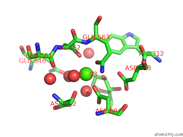
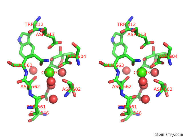
Calcium binding site 1 out of 4 in 7fe1
Go back to
Calcium binding site 1 out
of 4 in the Crystal Structure of GH92 Alpha-1,2-Mannosidase From Enterococcus Faecalis Atcc 10100 in Complex with Methyl Alpha-1,2-C-Mannobioside
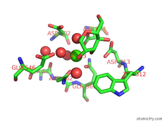
Mono view
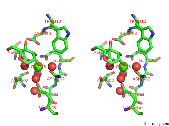
Stereo pair view
Mono view
Stereo pair view
A full contact list of Calcium with other atoms in the Ca binding
site number 1 of Crystal Structure of GH92 Alpha-1,2-Mannosidase From Enterococcus Faecalis Atcc 10100 in Complex with Methyl Alpha-1,2-C-Mannobioside within 5.0Å range:
|
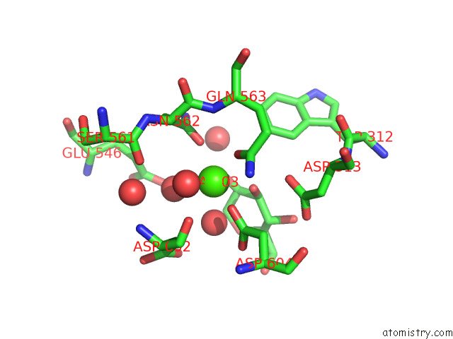
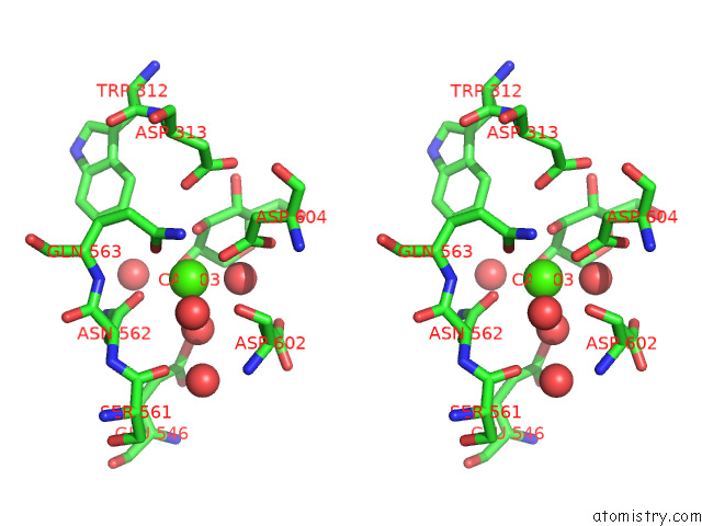
Calcium binding site 2 out of 4 in 7fe1
Go back to
Calcium binding site 2 out
of 4 in the Crystal Structure of GH92 Alpha-1,2-Mannosidase From Enterococcus Faecalis Atcc 10100 in Complex with Methyl Alpha-1,2-C-Mannobioside
Mono view
Stereo pair view
Mono view
Stereo pair view
A full contact list of Calcium with other atoms in the Ca binding
site number 2 of Crystal Structure of GH92 Alpha-1,2-Mannosidase From Enterococcus Faecalis Atcc 10100 in Complex with Methyl Alpha-1,2-C-Mannobioside within 5.0Å range:
|
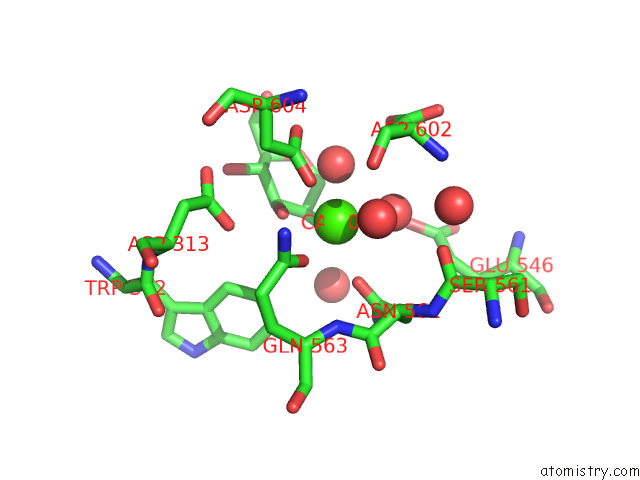
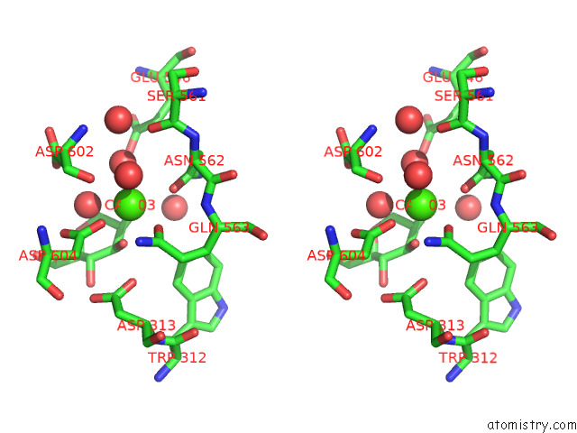
Calcium binding site 3 out of 4 in 7fe1
Go back to
Calcium binding site 3 out
of 4 in the Crystal Structure of GH92 Alpha-1,2-Mannosidase From Enterococcus Faecalis Atcc 10100 in Complex with Methyl Alpha-1,2-C-Mannobioside
Mono view
Stereo pair view
Mono view
Stereo pair view
A full contact list of Calcium with other atoms in the Ca binding
site number 3 of Crystal Structure of GH92 Alpha-1,2-Mannosidase From Enterococcus Faecalis Atcc 10100 in Complex with Methyl Alpha-1,2-C-Mannobioside within 5.0Å range:
|
Calcium binding site 4 out of 4 in 7fe1
Go back to
Calcium binding site 4 out
of 4 in the Crystal Structure of GH92 Alpha-1,2-Mannosidase From Enterococcus Faecalis Atcc 10100 in Complex with Methyl Alpha-1,2-C-Mannobioside
Mono view
Stereo pair view
Mono view
Stereo pair view
A full contact list of Calcium with other atoms in the Ca binding
site number 4 of Crystal Structure of GH92 Alpha-1,2-Mannosidase From Enterococcus Faecalis Atcc 10100 in Complex with Methyl Alpha-1,2-C-Mannobioside within 5.0Å range:
|
Reference:
S.Alonso-Gil,
K.Parkan,
J.Kaminsky,
R.Pohl,
T.Miyazaki.
Unlocking the Hydrolytic Mechanism of GH92 Alpha-1,2-Mannosidases: Computation Inspires the Use of C-Glycosides As Michaelis Complex Mimics. Chemistry V. 28 00148 2022.
ISSN: ISSN 0947-6539
PubMed: 35049087
DOI: 10.1002/CHEM.202200148
Page generated: Fri Jul 19 00:49:20 2024
ISSN: ISSN 0947-6539
PubMed: 35049087
DOI: 10.1002/CHEM.202200148
Last articles
Zn in 9J0NZn in 9J0O
Zn in 9J0P
Zn in 9FJX
Zn in 9EKB
Zn in 9C0F
Zn in 9CAH
Zn in 9CH0
Zn in 9CH3
Zn in 9CH1