Calcium »
PDB 7f9j-7g2r »
7ffp »
Calcium in PDB 7ffp: Crystal Structure of Di-Peptidase-E From Xenopus Laevis
Enzymatic activity of Crystal Structure of Di-Peptidase-E From Xenopus Laevis
All present enzymatic activity of Crystal Structure of Di-Peptidase-E From Xenopus Laevis:
3.4.13.21;
3.4.13.21;
Protein crystallography data
The structure of Crystal Structure of Di-Peptidase-E From Xenopus Laevis, PDB code: 7ffp
was solved by
A.Kumar,
R.Singh,
R.D.Makde,
with X-Ray Crystallography technique. A brief refinement statistics is given in the table below:
| Resolution Low / High (Å) | 29.22 / 1.80 |
| Space group | C 1 2 1 |
| Cell size a, b, c (Å), α, β, γ (°) | 82.666, 42.15, 75.68, 90, 118.24, 90 |
| R / Rfree (%) | 18.7 / 22.3 |
Calcium Binding Sites:
The binding sites of Calcium atom in the Crystal Structure of Di-Peptidase-E From Xenopus Laevis
(pdb code 7ffp). This binding sites where shown within
5.0 Angstroms radius around Calcium atom.
In total only one binding site of Calcium was determined in the Crystal Structure of Di-Peptidase-E From Xenopus Laevis, PDB code: 7ffp:
In total only one binding site of Calcium was determined in the Crystal Structure of Di-Peptidase-E From Xenopus Laevis, PDB code: 7ffp:
Calcium binding site 1 out of 1 in 7ffp
Go back to
Calcium binding site 1 out
of 1 in the Crystal Structure of Di-Peptidase-E From Xenopus Laevis
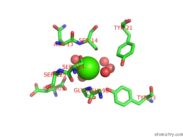
Mono view
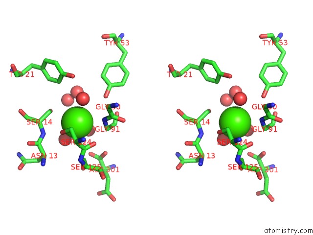
Stereo pair view
Mono view
Stereo pair view
A full contact list of Calcium with other atoms in the Ca binding
site number 1 of Crystal Structure of Di-Peptidase-E From Xenopus Laevis within 5.0Å range:
|
Reference:
A.Kumar,
R.Singh,
B.Ghosh,
R.D.Makde.
Crystal Structure of Aspartyl Dipeptidase From Xenopus Laevis Revealed Ligand Binding Induced Loop Ordering and Catalytic Triad Assembly. Proteins 2021.
ISSN: ESSN 1097-0134
PubMed: 34431561
DOI: 10.1002/PROT.26220
Page generated: Wed Jul 9 22:09:21 2025
ISSN: ESSN 1097-0134
PubMed: 34431561
DOI: 10.1002/PROT.26220
Last articles
Fe in 2YXOFe in 2YRS
Fe in 2YXC
Fe in 2YNM
Fe in 2YVJ
Fe in 2YP1
Fe in 2YU2
Fe in 2YU1
Fe in 2YQB
Fe in 2YOO