Calcium »
PDB 7g78-7jlg »
7jk1 »
Calcium in PDB 7jk1: Human Primpol Inserting Correct Dctp Opposite the 8-Oxoguanine Lesion
Protein crystallography data
The structure of Human Primpol Inserting Correct Dctp Opposite the 8-Oxoguanine Lesion, PDB code: 7jk1
was solved by
O.Rechkoblit,
A.K.Aggarwal,
with X-Ray Crystallography technique. A brief refinement statistics is given in the table below:
| Resolution Low / High (Å) | 39.88 / 2.62 |
| Space group | P 1 |
| Cell size a, b, c (Å), α, β, γ (°) | 52.558, 65.732, 75.179, 69.66, 82.83, 88.54 |
| R / Rfree (%) | 23.9 / 28.6 |
Calcium Binding Sites:
The binding sites of Calcium atom in the Human Primpol Inserting Correct Dctp Opposite the 8-Oxoguanine Lesion
(pdb code 7jk1). This binding sites where shown within
5.0 Angstroms radius around Calcium atom.
In total 2 binding sites of Calcium where determined in the Human Primpol Inserting Correct Dctp Opposite the 8-Oxoguanine Lesion, PDB code: 7jk1:
Jump to Calcium binding site number: 1; 2;
In total 2 binding sites of Calcium where determined in the Human Primpol Inserting Correct Dctp Opposite the 8-Oxoguanine Lesion, PDB code: 7jk1:
Jump to Calcium binding site number: 1; 2;
Calcium binding site 1 out of 2 in 7jk1
Go back to
Calcium binding site 1 out
of 2 in the Human Primpol Inserting Correct Dctp Opposite the 8-Oxoguanine Lesion
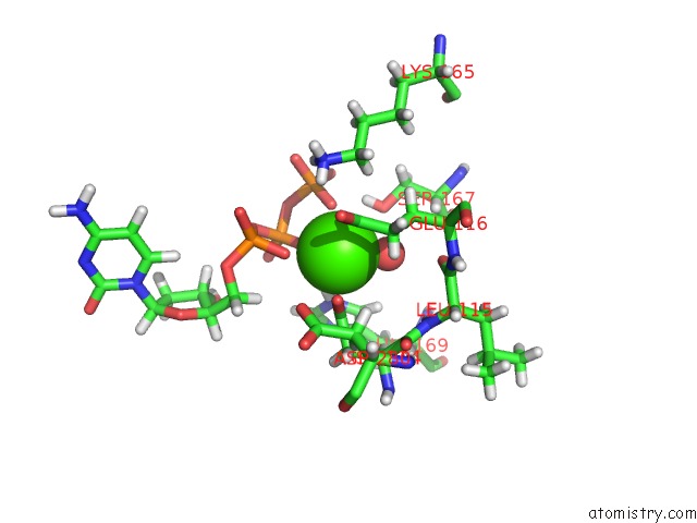
Mono view
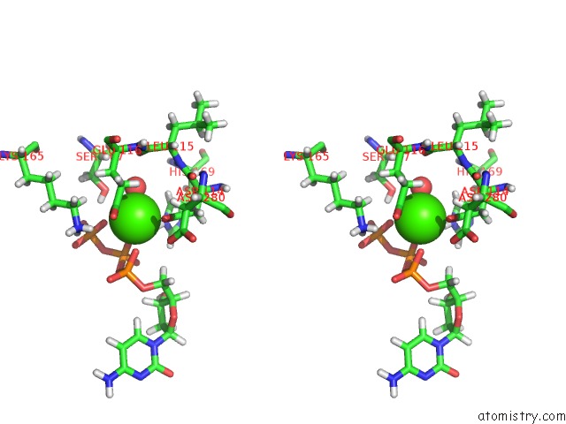
Stereo pair view
Mono view
Stereo pair view
A full contact list of Calcium with other atoms in the Ca binding
site number 1 of Human Primpol Inserting Correct Dctp Opposite the 8-Oxoguanine Lesion within 5.0Å range:
|
Calcium binding site 2 out of 2 in 7jk1
Go back to
Calcium binding site 2 out
of 2 in the Human Primpol Inserting Correct Dctp Opposite the 8-Oxoguanine Lesion
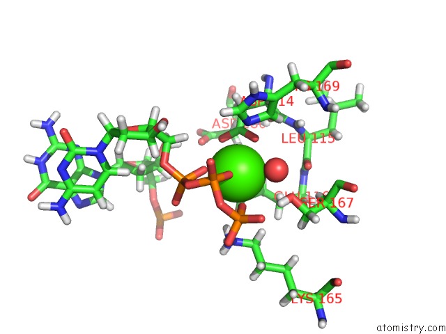
Mono view
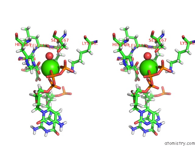
Stereo pair view
Mono view
Stereo pair view
A full contact list of Calcium with other atoms in the Ca binding
site number 2 of Human Primpol Inserting Correct Dctp Opposite the 8-Oxoguanine Lesion within 5.0Å range:
|
Reference:
O.Rechkoblit,
R.E.Johnson,
Y.K.Gupta,
L.Prakash,
S.Prakash,
A.K.Aggarwal.
Structural Basis of Dna Synthesis Opposite 8-Oxoguanine By Human Primpol Primase-Polymerase Nat Commun 2021.
ISSN: ESSN 2041-1723
DOI: 10.1038/S41467-021-24317-Z
Page generated: Wed Jul 9 22:41:47 2025
ISSN: ESSN 2041-1723
DOI: 10.1038/S41467-021-24317-Z
Last articles
Fe in 2YXOFe in 2YRS
Fe in 2YXC
Fe in 2YNM
Fe in 2YVJ
Fe in 2YP1
Fe in 2YU2
Fe in 2YU1
Fe in 2YQB
Fe in 2YOO