Calcium »
PDB 7g78-7jlg »
7jl8 »
Calcium in PDB 7jl8: Human Primpol Extending From the Correct Primer Base C Opposite the 8- Oxoguanine Lesion
Protein crystallography data
The structure of Human Primpol Extending From the Correct Primer Base C Opposite the 8- Oxoguanine Lesion, PDB code: 7jl8
was solved by
O.Rechkoblit,
A.K.Aggarwal,
with X-Ray Crystallography technique. A brief refinement statistics is given in the table below:
| Resolution Low / High (Å) | 28.16 / 2.10 |
| Space group | P 1 |
| Cell size a, b, c (Å), α, β, γ (°) | 50.689, 65.33, 72.001, 68.47, 85.67, 86.92 |
| R / Rfree (%) | 18.7 / 21.3 |
Calcium Binding Sites:
The binding sites of Calcium atom in the Human Primpol Extending From the Correct Primer Base C Opposite the 8- Oxoguanine Lesion
(pdb code 7jl8). This binding sites where shown within
5.0 Angstroms radius around Calcium atom.
In total 3 binding sites of Calcium where determined in the Human Primpol Extending From the Correct Primer Base C Opposite the 8- Oxoguanine Lesion, PDB code: 7jl8:
Jump to Calcium binding site number: 1; 2; 3;
In total 3 binding sites of Calcium where determined in the Human Primpol Extending From the Correct Primer Base C Opposite the 8- Oxoguanine Lesion, PDB code: 7jl8:
Jump to Calcium binding site number: 1; 2; 3;
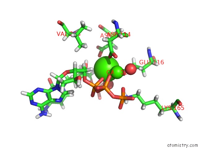
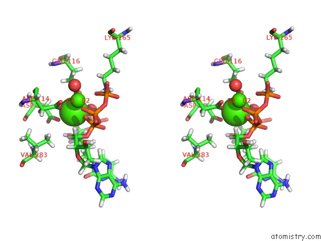
Calcium binding site 1 out of 3 in 7jl8
Go back to
Calcium binding site 1 out
of 3 in the Human Primpol Extending From the Correct Primer Base C Opposite the 8- Oxoguanine Lesion
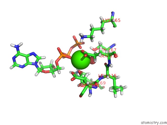
Mono view
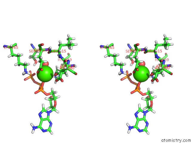
Stereo pair view
Mono view
Stereo pair view
A full contact list of Calcium with other atoms in the Ca binding
site number 1 of Human Primpol Extending From the Correct Primer Base C Opposite the 8- Oxoguanine Lesion within 5.0Å range:
|
Calcium binding site 2 out of 3 in 7jl8
Go back to
Calcium binding site 2 out
of 3 in the Human Primpol Extending From the Correct Primer Base C Opposite the 8- Oxoguanine Lesion
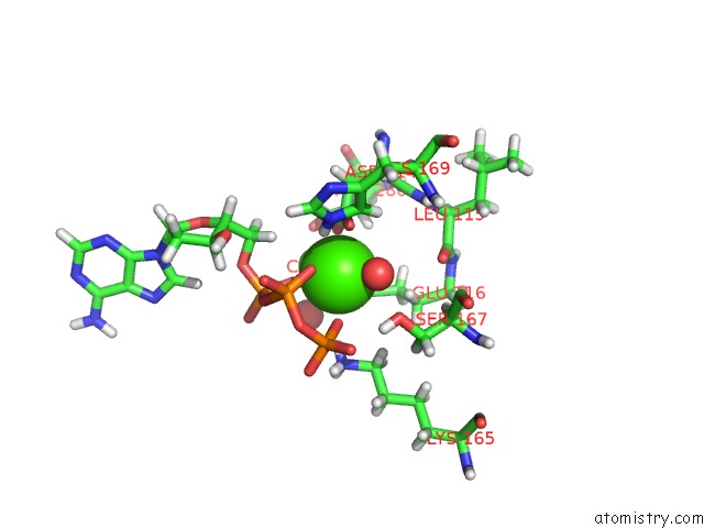
Mono view
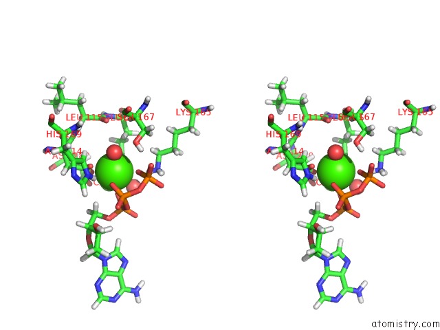
Stereo pair view
Mono view
Stereo pair view
A full contact list of Calcium with other atoms in the Ca binding
site number 2 of Human Primpol Extending From the Correct Primer Base C Opposite the 8- Oxoguanine Lesion within 5.0Å range:
|
Calcium binding site 3 out of 3 in 7jl8
Go back to
Calcium binding site 3 out
of 3 in the Human Primpol Extending From the Correct Primer Base C Opposite the 8- Oxoguanine Lesion
Mono view
Stereo pair view
Mono view
Stereo pair view
A full contact list of Calcium with other atoms in the Ca binding
site number 3 of Human Primpol Extending From the Correct Primer Base C Opposite the 8- Oxoguanine Lesion within 5.0Å range:
|
Reference:
O.Rechkoblit,
R.E.Johnson,
Y.K.Gupta,
L.Prakash,
S.Prakash,
A.K.Aggarwal.
Structural Basis of Dna Synthesis Opposite 8-Oxoguanine By Human Primpol Primase-Polymerase Nat Commun 2021.
ISSN: ESSN 2041-1723
DOI: 10.1038/S41467-021-24317-Z
Page generated: Wed Jul 9 22:42:20 2025
ISSN: ESSN 2041-1723
DOI: 10.1038/S41467-021-24317-Z
Last articles
F in 4KUWF in 4KOE
F in 4KSG
F in 4KQV
F in 4KS8
F in 4KNB
F in 4KRA
F in 4KPF
F in 4KPE
F in 4KN6