Calcium »
PDB 7jlu-7k04 »
7jvl »
Calcium in PDB 7jvl: Structure of the M101A Variant of the Sida Ornithine Hydroxylase Complexed with Nadp and the Fad in the "Out" Conformation
Enzymatic activity of Structure of the M101A Variant of the Sida Ornithine Hydroxylase Complexed with Nadp and the Fad in the "Out" Conformation
All present enzymatic activity of Structure of the M101A Variant of the Sida Ornithine Hydroxylase Complexed with Nadp and the Fad in the "Out" Conformation:
1.14.13.196;
1.14.13.196;
Protein crystallography data
The structure of Structure of the M101A Variant of the Sida Ornithine Hydroxylase Complexed with Nadp and the Fad in the "Out" Conformation, PDB code: 7jvl
was solved by
J.J.Tanner,
A.C.Campbell,
with X-Ray Crystallography technique. A brief refinement statistics is given in the table below:
| Resolution Low / High (Å) | 62.89 / 2.10 |
| Space group | P 1 21 1 |
| Cell size a, b, c (Å), α, β, γ (°) | 80.229, 153.811, 89.870, 90.00, 109.28, 90.00 |
| R / Rfree (%) | 20.4 / 24.1 |
Calcium Binding Sites:
The binding sites of Calcium atom in the Structure of the M101A Variant of the Sida Ornithine Hydroxylase Complexed with Nadp and the Fad in the "Out" Conformation
(pdb code 7jvl). This binding sites where shown within
5.0 Angstroms radius around Calcium atom.
In total 2 binding sites of Calcium where determined in the Structure of the M101A Variant of the Sida Ornithine Hydroxylase Complexed with Nadp and the Fad in the "Out" Conformation, PDB code: 7jvl:
Jump to Calcium binding site number: 1; 2;
In total 2 binding sites of Calcium where determined in the Structure of the M101A Variant of the Sida Ornithine Hydroxylase Complexed with Nadp and the Fad in the "Out" Conformation, PDB code: 7jvl:
Jump to Calcium binding site number: 1; 2;
Calcium binding site 1 out of 2 in 7jvl
Go back to
Calcium binding site 1 out
of 2 in the Structure of the M101A Variant of the Sida Ornithine Hydroxylase Complexed with Nadp and the Fad in the "Out" Conformation
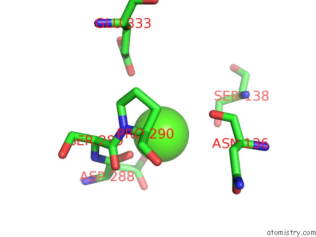
Mono view
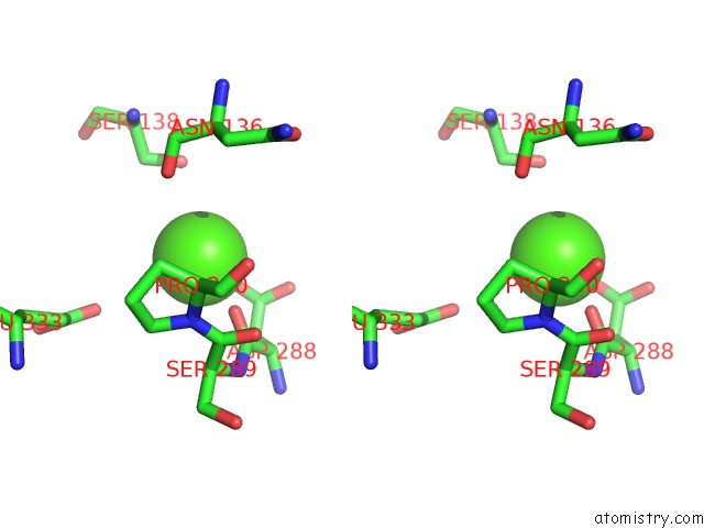
Stereo pair view
Mono view
Stereo pair view
A full contact list of Calcium with other atoms in the Ca binding
site number 1 of Structure of the M101A Variant of the Sida Ornithine Hydroxylase Complexed with Nadp and the Fad in the "Out" Conformation within 5.0Å range:
|
Calcium binding site 2 out of 2 in 7jvl
Go back to
Calcium binding site 2 out
of 2 in the Structure of the M101A Variant of the Sida Ornithine Hydroxylase Complexed with Nadp and the Fad in the "Out" Conformation
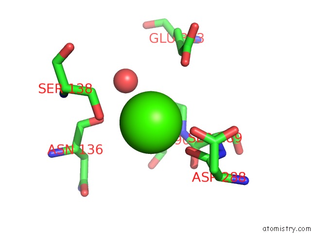
Mono view
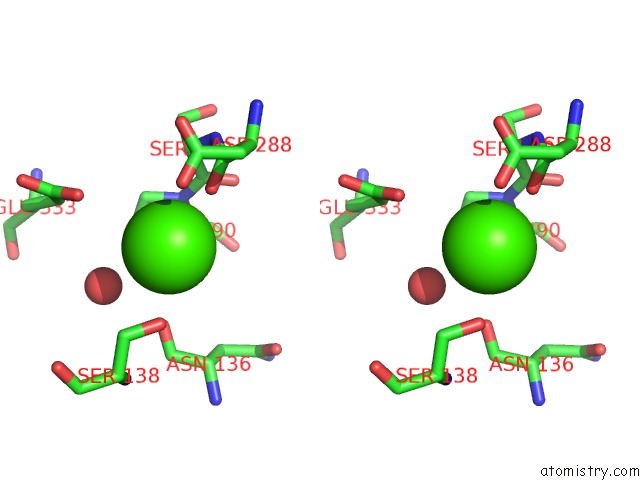
Stereo pair view
Mono view
Stereo pair view
A full contact list of Calcium with other atoms in the Ca binding
site number 2 of Structure of the M101A Variant of the Sida Ornithine Hydroxylase Complexed with Nadp and the Fad in the "Out" Conformation within 5.0Å range:
|
Reference:
A.C.Campbell,
R.Robinson,
D.Mena-Aguilar,
P.Sobrado,
J.J.Tanner.
Structural Determinants of Flavin Dynamics in A Class B Monooxygenase. Biochemistry 2020.
ISSN: ISSN 0006-2960
PubMed: 33226785
DOI: 10.1021/ACS.BIOCHEM.0C00783
Page generated: Wed Jul 9 22:48:59 2025
ISSN: ISSN 0006-2960
PubMed: 33226785
DOI: 10.1021/ACS.BIOCHEM.0C00783
Last articles
Fe in 2YXOFe in 2YRS
Fe in 2YXC
Fe in 2YNM
Fe in 2YVJ
Fe in 2YP1
Fe in 2YU2
Fe in 2YU1
Fe in 2YQB
Fe in 2YOO