Calcium »
PDB 7jlu-7k04 »
7jwf »
Calcium in PDB 7jwf: Crystal Structure of PDGH110B D344N in Complex with Alpha-(1,3)- Galactobiose
Protein crystallography data
The structure of Crystal Structure of PDGH110B D344N in Complex with Alpha-(1,3)- Galactobiose, PDB code: 7jwf
was solved by
A.G.Hettle,
A.B.Boraston,
with X-Ray Crystallography technique. A brief refinement statistics is given in the table below:
| Resolution Low / High (Å) | 29.76 / 2.19 |
| Space group | P 1 21 1 |
| Cell size a, b, c (Å), α, β, γ (°) | 98.517, 124.763, 142.423, 90.00, 93.86, 90.00 |
| R / Rfree (%) | 20.5 / 23.7 |
Other elements in 7jwf:
The structure of Crystal Structure of PDGH110B D344N in Complex with Alpha-(1,3)- Galactobiose also contains other interesting chemical elements:
| Iodine | (I) | 120 atoms |
| Chlorine | (Cl) | 10 atoms |
Calcium Binding Sites:
The binding sites of Calcium atom in the Crystal Structure of PDGH110B D344N in Complex with Alpha-(1,3)- Galactobiose
(pdb code 7jwf). This binding sites where shown within
5.0 Angstroms radius around Calcium atom.
In total only one binding site of Calcium was determined in the Crystal Structure of PDGH110B D344N in Complex with Alpha-(1,3)- Galactobiose, PDB code: 7jwf:
In total only one binding site of Calcium was determined in the Crystal Structure of PDGH110B D344N in Complex with Alpha-(1,3)- Galactobiose, PDB code: 7jwf:
Calcium binding site 1 out of 1 in 7jwf
Go back to
Calcium binding site 1 out
of 1 in the Crystal Structure of PDGH110B D344N in Complex with Alpha-(1,3)- Galactobiose
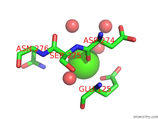
Mono view
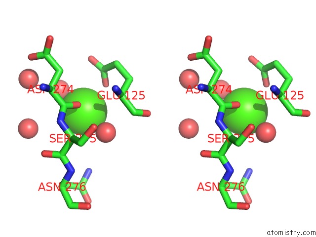
Stereo pair view
Mono view
Stereo pair view
A full contact list of Calcium with other atoms in the Ca binding
site number 1 of Crystal Structure of PDGH110B D344N in Complex with Alpha-(1,3)- Galactobiose within 5.0Å range:
|
Reference:
B.E.Mcguire,
A.Hettle,
C.Vickers,
D.T.King,
D.J.Vocadlo,
A.B.Boraston.
The Structure of A Family 110 Glycoside Hydrolase Provides Insight Into the Hydrolysis of Alpha-(1,3)-Galactosidic Linkages in Lambda-Carrageenan and Blood Group Antigens. J.Biol.Chem. 2020.
ISSN: ESSN 1083-351X
PubMed: 33127644
DOI: 10.1074/JBC.RA120.015776
Page generated: Wed Jul 9 22:49:15 2025
ISSN: ESSN 1083-351X
PubMed: 33127644
DOI: 10.1074/JBC.RA120.015776
Last articles
Fe in 2YXOFe in 2YRS
Fe in 2YXC
Fe in 2YNM
Fe in 2YVJ
Fe in 2YP1
Fe in 2YU2
Fe in 2YU1
Fe in 2YQB
Fe in 2YOO