Calcium »
PDB 7k1r-7kl5 »
7k1r »
Calcium in PDB 7k1r: X-Ray Structure of An Enterobacter GH43 Beta-Xylosidase: ECXYL43 F507A Mutant
Enzymatic activity of X-Ray Structure of An Enterobacter GH43 Beta-Xylosidase: ECXYL43 F507A Mutant
All present enzymatic activity of X-Ray Structure of An Enterobacter GH43 Beta-Xylosidase: ECXYL43 F507A Mutant:
3.2.1.37;
3.2.1.37;
Protein crystallography data
The structure of X-Ray Structure of An Enterobacter GH43 Beta-Xylosidase: ECXYL43 F507A Mutant, PDB code: 7k1r
was solved by
L.Briganti,
C.C.M.Capetti,
I.Polikarpov,
with X-Ray Crystallography technique. A brief refinement statistics is given in the table below:
| Resolution Low / High (Å) | 22.72 / 2.40 |
| Space group | P 21 21 21 |
| Cell size a, b, c (Å), α, β, γ (°) | 87.123, 153.1, 182.401, 90, 90, 90 |
| R / Rfree (%) | 19 / 22.8 |
Calcium Binding Sites:
The binding sites of Calcium atom in the X-Ray Structure of An Enterobacter GH43 Beta-Xylosidase: ECXYL43 F507A Mutant
(pdb code 7k1r). This binding sites where shown within
5.0 Angstroms radius around Calcium atom.
In total 4 binding sites of Calcium where determined in the X-Ray Structure of An Enterobacter GH43 Beta-Xylosidase: ECXYL43 F507A Mutant, PDB code: 7k1r:
Jump to Calcium binding site number: 1; 2; 3; 4;
In total 4 binding sites of Calcium where determined in the X-Ray Structure of An Enterobacter GH43 Beta-Xylosidase: ECXYL43 F507A Mutant, PDB code: 7k1r:
Jump to Calcium binding site number: 1; 2; 3; 4;
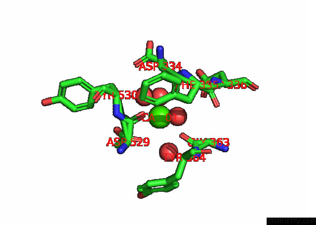
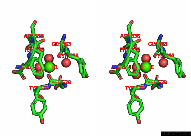
Calcium binding site 1 out of 4 in 7k1r
Go back to
Calcium binding site 1 out
of 4 in the X-Ray Structure of An Enterobacter GH43 Beta-Xylosidase: ECXYL43 F507A Mutant
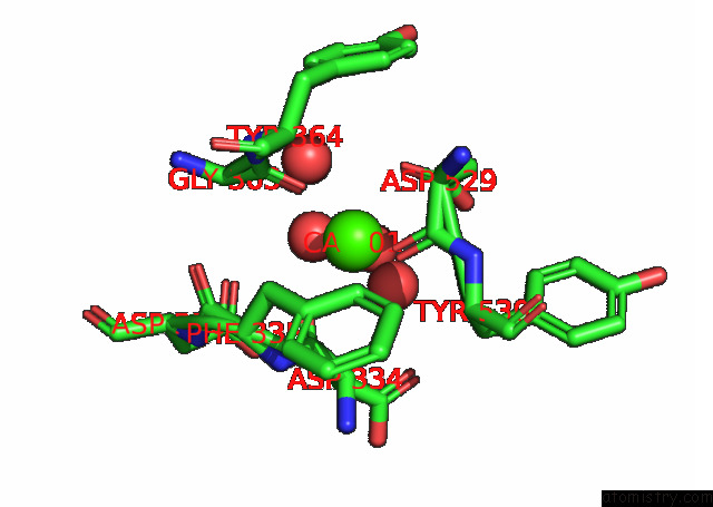
Mono view
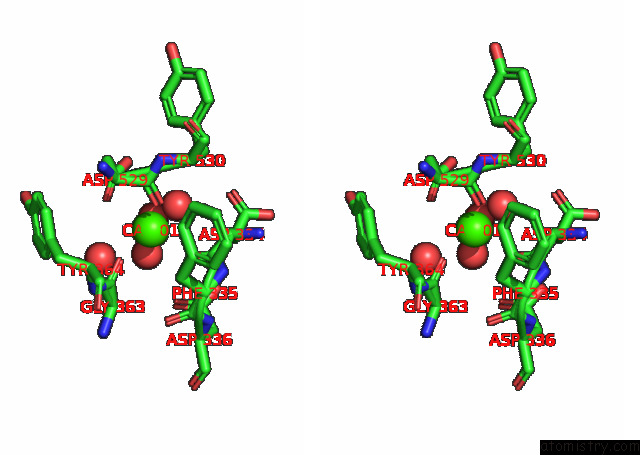
Stereo pair view
Mono view
Stereo pair view
A full contact list of Calcium with other atoms in the Ca binding
site number 1 of X-Ray Structure of An Enterobacter GH43 Beta-Xylosidase: ECXYL43 F507A Mutant within 5.0Å range:
|
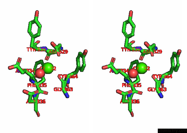
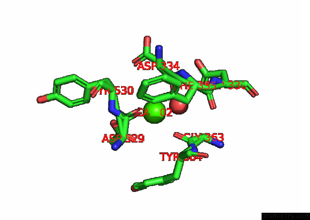
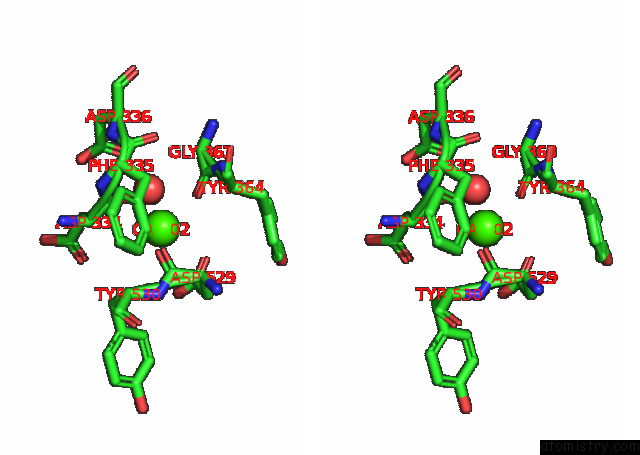
Calcium binding site 2 out of 4 in 7k1r
Go back to
Calcium binding site 2 out
of 4 in the X-Ray Structure of An Enterobacter GH43 Beta-Xylosidase: ECXYL43 F507A Mutant
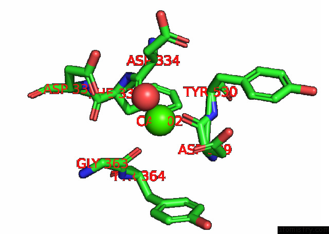
Mono view
Stereo pair view
Mono view
Stereo pair view
A full contact list of Calcium with other atoms in the Ca binding
site number 2 of X-Ray Structure of An Enterobacter GH43 Beta-Xylosidase: ECXYL43 F507A Mutant within 5.0Å range:
|
Calcium binding site 3 out of 4 in 7k1r
Go back to
Calcium binding site 3 out
of 4 in the X-Ray Structure of An Enterobacter GH43 Beta-Xylosidase: ECXYL43 F507A Mutant
Mono view
Stereo pair view
Mono view
Stereo pair view
A full contact list of Calcium with other atoms in the Ca binding
site number 3 of X-Ray Structure of An Enterobacter GH43 Beta-Xylosidase: ECXYL43 F507A Mutant within 5.0Å range:
|
Calcium binding site 4 out of 4 in 7k1r
Go back to
Calcium binding site 4 out
of 4 in the X-Ray Structure of An Enterobacter GH43 Beta-Xylosidase: ECXYL43 F507A Mutant
Mono view
Stereo pair view
Mono view
Stereo pair view
A full contact list of Calcium with other atoms in the Ca binding
site number 4 of X-Ray Structure of An Enterobacter GH43 Beta-Xylosidase: ECXYL43 F507A Mutant within 5.0Å range:
|
Reference:
L.Briganti,
C.C.M.Capetti,
I.Polikarpov.
X-Ray Structure of An Enterobacter GH43 Beta-Xylosidase: ECXYL43 F507A Mutant To Be Published.
Page generated: Wed Jul 9 22:49:57 2025
Last articles
Fe in 2YXOFe in 2YRS
Fe in 2YXC
Fe in 2YNM
Fe in 2YVJ
Fe in 2YP1
Fe in 2YU2
Fe in 2YU1
Fe in 2YQB
Fe in 2YOO