Calcium »
PDB 7k1r-7kl5 »
7k72 »
Calcium in PDB 7k72: Structure of Dna Ligase A From Mycobacterium Tuberculosis Bound to Nad
Enzymatic activity of Structure of Dna Ligase A From Mycobacterium Tuberculosis Bound to Nad
All present enzymatic activity of Structure of Dna Ligase A From Mycobacterium Tuberculosis Bound to Nad:
6.5.1.2;
6.5.1.2;
Protein crystallography data
The structure of Structure of Dna Ligase A From Mycobacterium Tuberculosis Bound to Nad, PDB code: 7k72
was solved by
Seattle Structural Genomics Center For Infectious Disease (Ssgcid),
with X-Ray Crystallography technique. A brief refinement statistics is given in the table below:
| Resolution Low / High (Å) | 44.59 / 2.05 |
| Space group | P 1 |
| Cell size a, b, c (Å), α, β, γ (°) | 50.940, 63.810, 120.730, 96.22, 89.55, 111.97 |
| R / Rfree (%) | 16.8 / 20.6 |
Other elements in 7k72:
The structure of Structure of Dna Ligase A From Mycobacterium Tuberculosis Bound to Nad also contains other interesting chemical elements:
| Magnesium | (Mg) | 9 atoms |
Calcium Binding Sites:
The binding sites of Calcium atom in the Structure of Dna Ligase A From Mycobacterium Tuberculosis Bound to Nad
(pdb code 7k72). This binding sites where shown within
5.0 Angstroms radius around Calcium atom.
In total 2 binding sites of Calcium where determined in the Structure of Dna Ligase A From Mycobacterium Tuberculosis Bound to Nad, PDB code: 7k72:
Jump to Calcium binding site number: 1; 2;
In total 2 binding sites of Calcium where determined in the Structure of Dna Ligase A From Mycobacterium Tuberculosis Bound to Nad, PDB code: 7k72:
Jump to Calcium binding site number: 1; 2;
Calcium binding site 1 out of 2 in 7k72
Go back to
Calcium binding site 1 out
of 2 in the Structure of Dna Ligase A From Mycobacterium Tuberculosis Bound to Nad
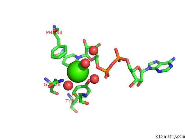
Mono view
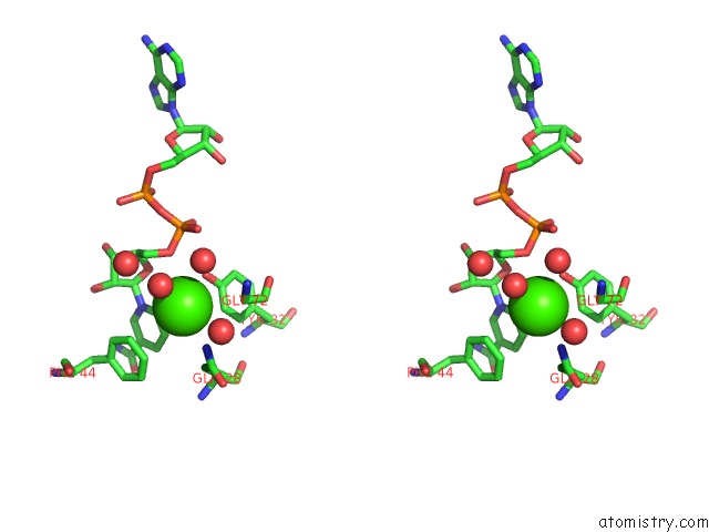
Stereo pair view
Mono view
Stereo pair view
A full contact list of Calcium with other atoms in the Ca binding
site number 1 of Structure of Dna Ligase A From Mycobacterium Tuberculosis Bound to Nad within 5.0Å range:
|
Calcium binding site 2 out of 2 in 7k72
Go back to
Calcium binding site 2 out
of 2 in the Structure of Dna Ligase A From Mycobacterium Tuberculosis Bound to Nad
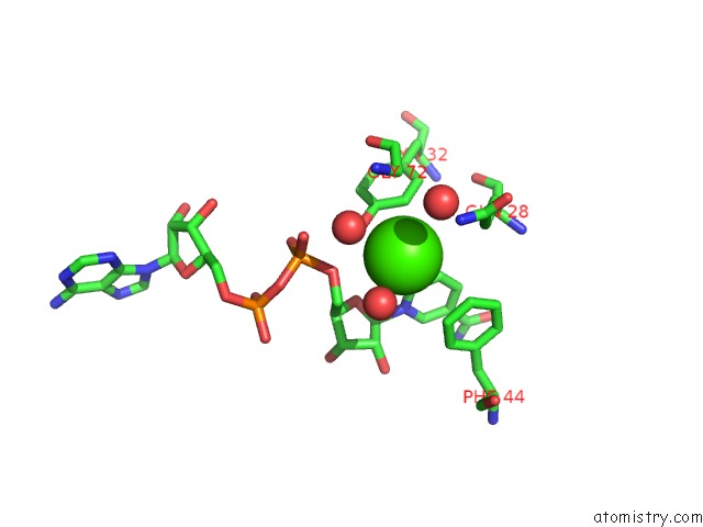
Mono view
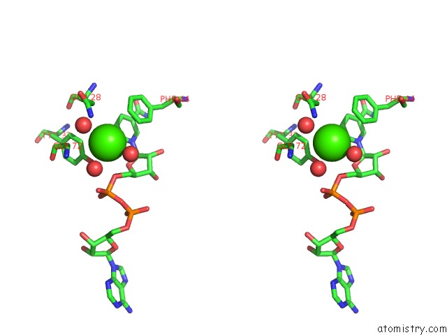
Stereo pair view
Mono view
Stereo pair view
A full contact list of Calcium with other atoms in the Ca binding
site number 2 of Structure of Dna Ligase A From Mycobacterium Tuberculosis Bound to Nad within 5.0Å range:
|
Reference:
S.L.Delker,
J.Abendroth,
D.D.Lorimer,
P.S.Horanyi,
T.E.Edwards.
Structure of Dna Ligase A From Mycobacterium Tuberculosis Bound to Nad To Be Published.
Page generated: Wed Jul 9 22:50:53 2025
Last articles
F in 7OHNF in 7OIH
F in 7OHL
F in 7OH6
F in 7OHK
F in 7OHH
F in 7OH5
F in 7OFK
F in 7OFI
F in 7OAR