Calcium »
PDB 7k1r-7kl5 »
7k9t »
Calcium in PDB 7k9t: Co-Crystal Structure of Alpha Glucosidase with Compound 5
Protein crystallography data
The structure of Co-Crystal Structure of Alpha Glucosidase with Compound 5, PDB code: 7k9t
was solved by
S.S.Karade,
R.A.Mariuzza,
with X-Ray Crystallography technique. A brief refinement statistics is given in the table below:
| Resolution Low / High (Å) | 42.35 / 2.10 |
| Space group | P 32 |
| Cell size a, b, c (Å), α, β, γ (°) | 102.87, 102.87, 240.653, 90, 90, 120 |
| R / Rfree (%) | 17.1 / 19.2 |
Calcium Binding Sites:
The binding sites of Calcium atom in the Co-Crystal Structure of Alpha Glucosidase with Compound 5
(pdb code 7k9t). This binding sites where shown within
5.0 Angstroms radius around Calcium atom.
In total 4 binding sites of Calcium where determined in the Co-Crystal Structure of Alpha Glucosidase with Compound 5, PDB code: 7k9t:
Jump to Calcium binding site number: 1; 2; 3; 4;
In total 4 binding sites of Calcium where determined in the Co-Crystal Structure of Alpha Glucosidase with Compound 5, PDB code: 7k9t:
Jump to Calcium binding site number: 1; 2; 3; 4;
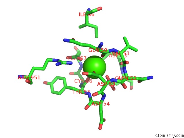
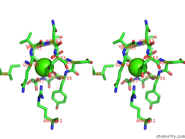
Calcium binding site 1 out of 4 in 7k9t
Go back to
Calcium binding site 1 out
of 4 in the Co-Crystal Structure of Alpha Glucosidase with Compound 5
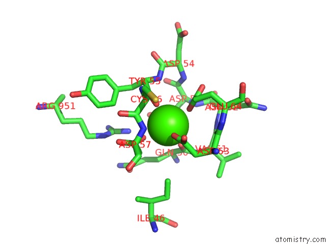
Mono view
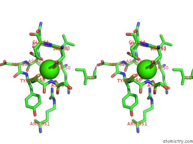
Stereo pair view
Mono view
Stereo pair view
A full contact list of Calcium with other atoms in the Ca binding
site number 1 of Co-Crystal Structure of Alpha Glucosidase with Compound 5 within 5.0Å range:
|
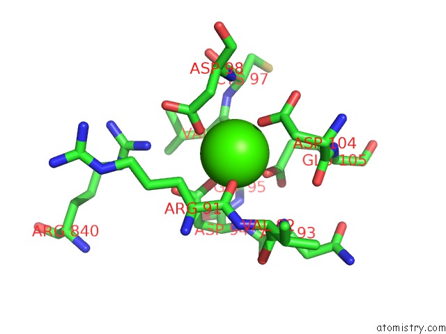
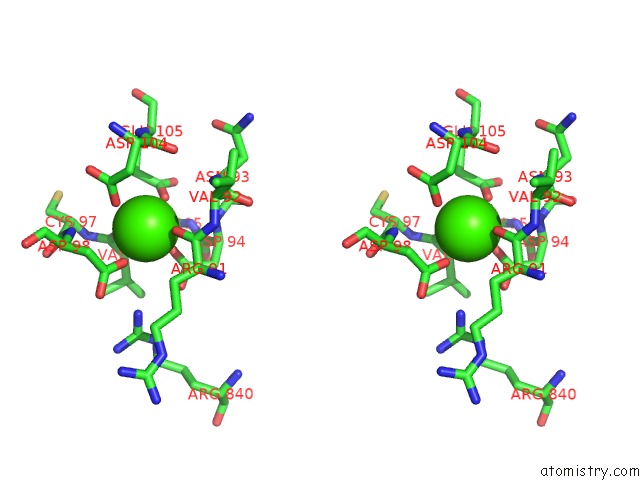
Calcium binding site 2 out of 4 in 7k9t
Go back to
Calcium binding site 2 out
of 4 in the Co-Crystal Structure of Alpha Glucosidase with Compound 5
Mono view
Stereo pair view
Mono view
Stereo pair view
A full contact list of Calcium with other atoms in the Ca binding
site number 2 of Co-Crystal Structure of Alpha Glucosidase with Compound 5 within 5.0Å range:
|
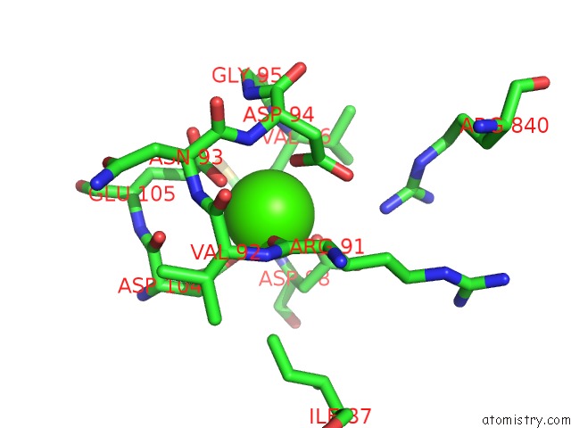
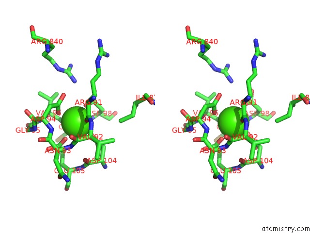
Calcium binding site 3 out of 4 in 7k9t
Go back to
Calcium binding site 3 out
of 4 in the Co-Crystal Structure of Alpha Glucosidase with Compound 5
Mono view
Stereo pair view
Mono view
Stereo pair view
A full contact list of Calcium with other atoms in the Ca binding
site number 3 of Co-Crystal Structure of Alpha Glucosidase with Compound 5 within 5.0Å range:
|
Calcium binding site 4 out of 4 in 7k9t
Go back to
Calcium binding site 4 out
of 4 in the Co-Crystal Structure of Alpha Glucosidase with Compound 5
Mono view
Stereo pair view
Mono view
Stereo pair view
A full contact list of Calcium with other atoms in the Ca binding
site number 4 of Co-Crystal Structure of Alpha Glucosidase with Compound 5 within 5.0Å range:
|
Reference:
S.S.Karade,
R.A.Mariuzza.
Co-Crystal Structure of Alpha Glucosidase with Compound 5 To Be Published.
Page generated: Wed Jul 9 22:51:43 2025
Last articles
F in 7Q2YF in 7Q3B
F in 7Q2X
F in 7PVK
F in 7Q2J
F in 7Q01
F in 7PZX
F in 7PZW
F in 7PZV
F in 7PZU