Calcium »
PDB 7kld-7l6g »
7kle »
Calcium in PDB 7kle: Ternary Structure of DPO4 Bound to N7MG in the Template Base Paired with Incoming Dctp
Enzymatic activity of Ternary Structure of DPO4 Bound to N7MG in the Template Base Paired with Incoming Dctp
All present enzymatic activity of Ternary Structure of DPO4 Bound to N7MG in the Template Base Paired with Incoming Dctp:
2.7.7.7;
2.7.7.7;
Protein crystallography data
The structure of Ternary Structure of DPO4 Bound to N7MG in the Template Base Paired with Incoming Dctp, PDB code: 7kle
was solved by
M.-C.Koag,
S.Lee,
with X-Ray Crystallography technique. A brief refinement statistics is given in the table below:
| Resolution Low / High (Å) | 19.56 / 3.00 |
| Space group | P 21 21 2 |
| Cell size a, b, c (Å), α, β, γ (°) | 99.65, 102.472, 53.034, 90, 90, 90 |
| R / Rfree (%) | 22.9 / 28.8 |
Other elements in 7kle:
The structure of Ternary Structure of DPO4 Bound to N7MG in the Template Base Paired with Incoming Dctp also contains other interesting chemical elements:
| Fluorine | (F) | 1 atom |
Calcium Binding Sites:
The binding sites of Calcium atom in the Ternary Structure of DPO4 Bound to N7MG in the Template Base Paired with Incoming Dctp
(pdb code 7kle). This binding sites where shown within
5.0 Angstroms radius around Calcium atom.
In total 3 binding sites of Calcium where determined in the Ternary Structure of DPO4 Bound to N7MG in the Template Base Paired with Incoming Dctp, PDB code: 7kle:
Jump to Calcium binding site number: 1; 2; 3;
In total 3 binding sites of Calcium where determined in the Ternary Structure of DPO4 Bound to N7MG in the Template Base Paired with Incoming Dctp, PDB code: 7kle:
Jump to Calcium binding site number: 1; 2; 3;
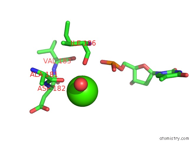
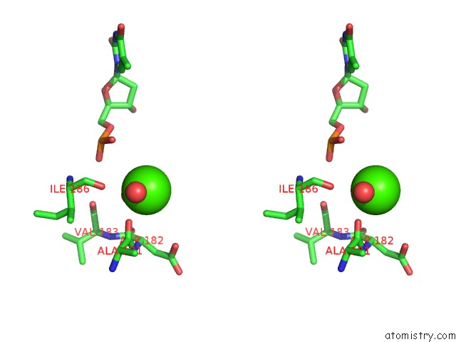
Calcium binding site 1 out of 3 in 7kle
Go back to
Calcium binding site 1 out
of 3 in the Ternary Structure of DPO4 Bound to N7MG in the Template Base Paired with Incoming Dctp
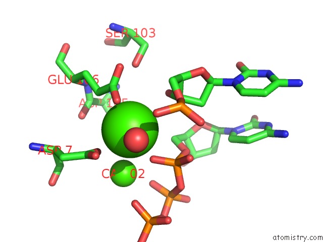
Mono view
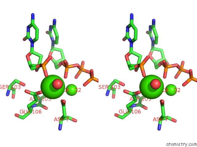
Stereo pair view
Mono view
Stereo pair view
A full contact list of Calcium with other atoms in the Ca binding
site number 1 of Ternary Structure of DPO4 Bound to N7MG in the Template Base Paired with Incoming Dctp within 5.0Å range:
|
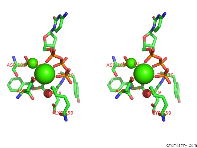
Calcium binding site 2 out of 3 in 7kle
Go back to
Calcium binding site 2 out
of 3 in the Ternary Structure of DPO4 Bound to N7MG in the Template Base Paired with Incoming Dctp
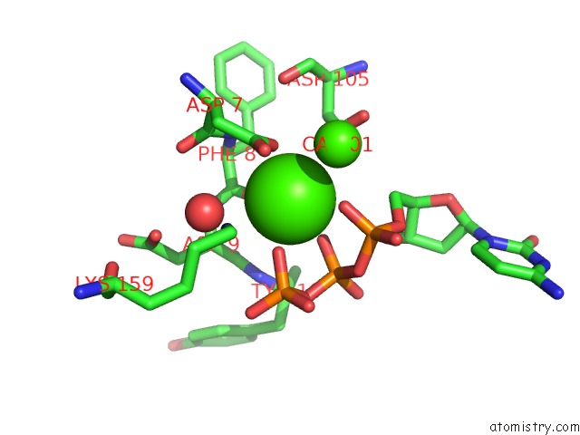
Mono view
Stereo pair view
Mono view
Stereo pair view
A full contact list of Calcium with other atoms in the Ca binding
site number 2 of Ternary Structure of DPO4 Bound to N7MG in the Template Base Paired with Incoming Dctp within 5.0Å range:
|
Calcium binding site 3 out of 3 in 7kle
Go back to
Calcium binding site 3 out
of 3 in the Ternary Structure of DPO4 Bound to N7MG in the Template Base Paired with Incoming Dctp
Mono view
Stereo pair view
Mono view
Stereo pair view
A full contact list of Calcium with other atoms in the Ca binding
site number 3 of Ternary Structure of DPO4 Bound to N7MG in the Template Base Paired with Incoming Dctp within 5.0Å range:
|
Reference:
M.-C.Koag,
S.Lee.
Structure of Human Dna Polymerase Beta Complexed with 8OA in the Template Base Paired with Incoming Non-Hydrolyzable Atp To Be Published.
Page generated: Wed Jul 9 22:57:40 2025
Last articles
Fe in 2YXOFe in 2YRS
Fe in 2YXC
Fe in 2YNM
Fe in 2YVJ
Fe in 2YP1
Fe in 2YU2
Fe in 2YU1
Fe in 2YQB
Fe in 2YOO