Calcium in PDB 7knv: Solution uc(Nmr) Structure of CDHR3 Extracellular Domain EC1
Calcium Binding Sites:
The binding sites of Calcium atom in the Solution uc(Nmr) Structure of CDHR3 Extracellular Domain EC1
(pdb code 7knv). This binding sites where shown within
5.0 Angstroms radius around Calcium atom.
In total 3 binding sites of Calcium where determined in the
Solution uc(Nmr) Structure of CDHR3 Extracellular Domain EC1, PDB code: 7knv:
Jump to Calcium binding site number:
1;
2;
3;
Calcium binding site 1 out
of 3 in 7knv
Go back to
Calcium Binding Sites List in 7knv
Calcium binding site 1 out
of 3 in the Solution uc(Nmr) Structure of CDHR3 Extracellular Domain EC1
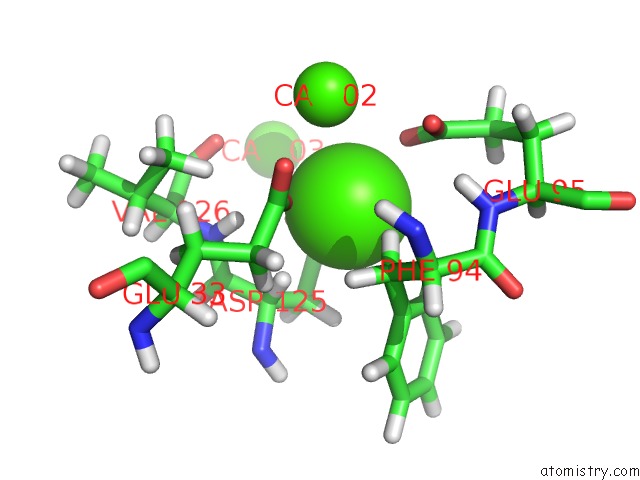
Mono view
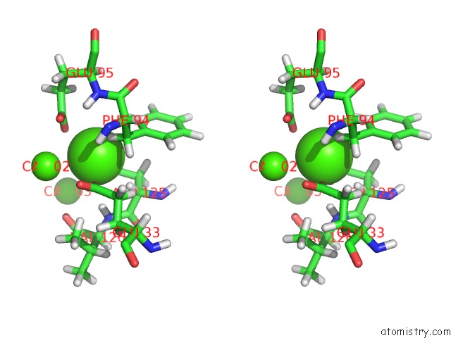
Stereo pair view
|
A full contact list of Calcium with other atoms in the Ca binding
site number 1 of Solution uc(Nmr) Structure of CDHR3 Extracellular Domain EC1 within 5.0Å range:
probe
|
atom
|
residue
|
distance (Å)
|
B
|
Occ
|
A:Ca201
b:0.0
occ:1.00
|
HB3
|
A:PHE94
|
2.1
|
0.0
|
1.0
|
OD1
|
A:ASP125
|
2.3
|
0.0
|
1.0
|
OE2
|
A:GLU33
|
2.3
|
0.0
|
1.0
|
CG
|
A:ASP125
|
2.5
|
0.0
|
1.0
|
OE2
|
A:GLU95
|
2.6
|
0.0
|
1.0
|
HA
|
A:ASP125
|
2.8
|
0.0
|
1.0
|
OD2
|
A:ASP125
|
2.9
|
0.0
|
1.0
|
CB
|
A:PHE94
|
3.0
|
0.0
|
1.0
|
HD2
|
A:PHE94
|
3.0
|
0.0
|
1.0
|
HB2
|
A:PHE94
|
3.1
|
0.0
|
1.0
|
H
|
A:VAL126
|
3.2
|
0.0
|
1.0
|
CD
|
A:GLU33
|
3.3
|
0.0
|
1.0
|
CA
|
A:CA202
|
3.3
|
0.0
|
1.0
|
CB
|
A:ASP125
|
3.4
|
0.0
|
1.0
|
CD
|
A:GLU95
|
3.4
|
0.0
|
1.0
|
HB2
|
A:ASP125
|
3.5
|
0.0
|
1.0
|
CG
|
A:PHE94
|
3.5
|
0.0
|
1.0
|
CD2
|
A:PHE94
|
3.5
|
0.0
|
1.0
|
CA
|
A:ASP125
|
3.5
|
0.0
|
1.0
|
OE1
|
A:GLU95
|
3.7
|
0.0
|
1.0
|
OE1
|
A:GLU33
|
3.9
|
0.0
|
1.0
|
N
|
A:VAL126
|
3.9
|
0.0
|
1.0
|
HG3
|
A:GLU33
|
4.0
|
0.0
|
1.0
|
H
|
A:GLU95
|
4.0
|
0.0
|
1.0
|
H
|
A:PHE94
|
4.0
|
0.0
|
1.0
|
CA
|
A:PHE94
|
4.2
|
0.0
|
1.0
|
C
|
A:ASP125
|
4.2
|
0.0
|
1.0
|
CG
|
A:GLU33
|
4.3
|
0.0
|
1.0
|
HG11
|
A:VAL126
|
4.3
|
0.0
|
1.0
|
HB3
|
A:ASP125
|
4.4
|
0.0
|
1.0
|
O
|
A:VAL126
|
4.5
|
0.0
|
1.0
|
N
|
A:GLU95
|
4.5
|
0.0
|
1.0
|
HG3
|
A:GLU95
|
4.5
|
0.0
|
1.0
|
N
|
A:PHE94
|
4.5
|
0.0
|
1.0
|
CG
|
A:GLU95
|
4.6
|
0.0
|
1.0
|
C
|
A:PHE94
|
4.6
|
0.0
|
1.0
|
CD1
|
A:PHE94
|
4.7
|
0.0
|
1.0
|
CE2
|
A:PHE94
|
4.7
|
0.0
|
1.0
|
N
|
A:ASP125
|
4.7
|
0.0
|
1.0
|
HG2
|
A:GLU33
|
4.8
|
0.0
|
1.0
|
CA
|
A:CA203
|
4.9
|
0.0
|
1.0
|
|
Calcium binding site 2 out
of 3 in 7knv
Go back to
Calcium Binding Sites List in 7knv
Calcium binding site 2 out
of 3 in the Solution uc(Nmr) Structure of CDHR3 Extracellular Domain EC1
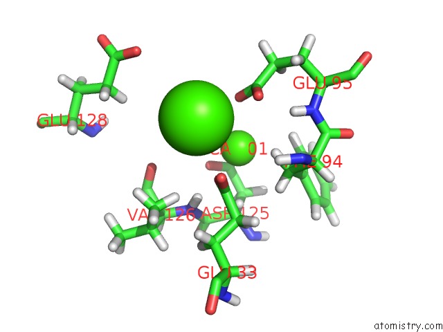
Mono view
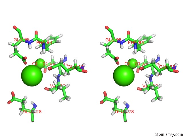
Stereo pair view
|
A full contact list of Calcium with other atoms in the Ca binding
site number 2 of Solution uc(Nmr) Structure of CDHR3 Extracellular Domain EC1 within 5.0Å range:
probe
|
atom
|
residue
|
distance (Å)
|
B
|
Occ
|
A:Ca202
b:0.0
occ:1.00
|
OE2
|
A:GLU95
|
2.8
|
0.0
|
1.0
|
OE1
|
A:GLU95
|
3.1
|
0.0
|
1.0
|
OE2
|
A:GLU33
|
3.2
|
0.0
|
1.0
|
CA
|
A:CA201
|
3.3
|
0.0
|
1.0
|
CD
|
A:GLU95
|
3.3
|
0.0
|
1.0
|
OE1
|
A:GLU33
|
3.4
|
0.0
|
1.0
|
CD
|
A:GLU33
|
3.7
|
0.0
|
1.0
|
HG11
|
A:VAL126
|
3.9
|
0.0
|
1.0
|
O
|
A:VAL126
|
4.1
|
0.0
|
1.0
|
HG3
|
A:GLU128
|
4.3
|
0.0
|
1.0
|
OE2
|
A:GLU128
|
4.3
|
0.0
|
1.0
|
HB3
|
A:PHE94
|
4.4
|
0.0
|
1.0
|
H
|
A:VAL126
|
4.4
|
0.0
|
1.0
|
OD1
|
A:ASP125
|
4.4
|
0.0
|
1.0
|
HG13
|
A:VAL126
|
4.5
|
0.0
|
1.0
|
HG2
|
A:GLU128
|
4.5
|
0.0
|
1.0
|
H
|
A:PHE94
|
4.6
|
0.0
|
1.0
|
H
|
A:GLU95
|
4.7
|
0.0
|
1.0
|
CG1
|
A:VAL126
|
4.7
|
0.0
|
1.0
|
CG
|
A:GLU95
|
4.8
|
0.0
|
1.0
|
CG
|
A:GLU128
|
4.8
|
0.0
|
1.0
|
HG2
|
A:GLU95
|
4.9
|
0.0
|
1.0
|
|
Calcium binding site 3 out
of 3 in 7knv
Go back to
Calcium Binding Sites List in 7knv
Calcium binding site 3 out
of 3 in the Solution uc(Nmr) Structure of CDHR3 Extracellular Domain EC1
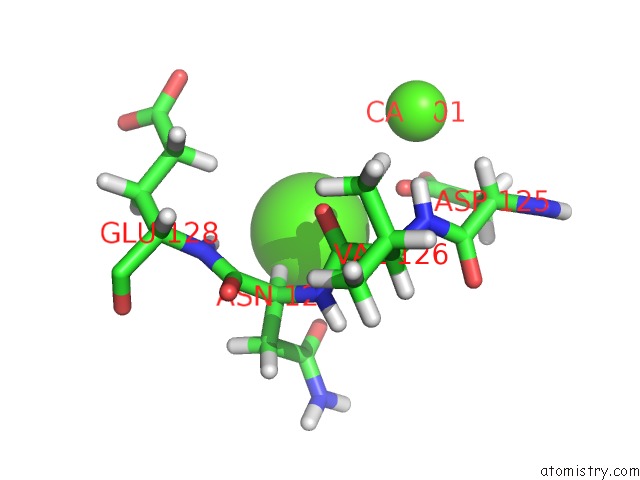
Mono view
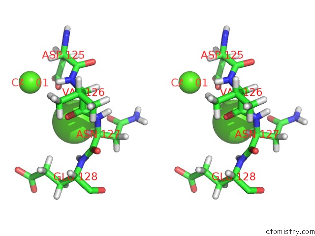
Stereo pair view
|
A full contact list of Calcium with other atoms in the Ca binding
site number 3 of Solution uc(Nmr) Structure of CDHR3 Extracellular Domain EC1 within 5.0Å range:
probe
|
atom
|
residue
|
distance (Å)
|
B
|
Occ
|
A:Ca203
b:0.0
occ:1.00
|
HA
|
A:ASN127
|
2.1
|
0.0
|
1.0
|
OD1
|
A:ASN127
|
2.9
|
0.0
|
1.0
|
CA
|
A:ASN127
|
3.1
|
0.0
|
1.0
|
O
|
A:VAL126
|
3.1
|
0.0
|
1.0
|
OD1
|
A:ASP125
|
3.2
|
0.0
|
1.0
|
H
|
A:GLU128
|
3.3
|
0.0
|
1.0
|
C
|
A:VAL126
|
3.5
|
0.0
|
1.0
|
N
|
A:ASN127
|
3.5
|
0.0
|
1.0
|
CG
|
A:ASN127
|
3.8
|
0.0
|
1.0
|
CG
|
A:ASP125
|
3.9
|
0.0
|
1.0
|
CB
|
A:ASN127
|
4.0
|
0.0
|
1.0
|
HG3
|
A:GLU128
|
4.0
|
0.0
|
1.0
|
N
|
A:GLU128
|
4.0
|
0.0
|
1.0
|
OD2
|
A:ASP125
|
4.0
|
0.0
|
1.0
|
C
|
A:ASN127
|
4.1
|
0.0
|
1.0
|
HB3
|
A:ASN127
|
4.2
|
0.0
|
1.0
|
H
|
A:ASN127
|
4.3
|
0.0
|
1.0
|
CA
|
A:VAL126
|
4.7
|
0.0
|
1.0
|
N
|
A:VAL126
|
4.7
|
0.0
|
1.0
|
H
|
A:VAL126
|
4.8
|
0.0
|
1.0
|
CA
|
A:CA201
|
4.9
|
0.0
|
1.0
|
HB2
|
A:ASN127
|
4.9
|
0.0
|
1.0
|
ND2
|
A:ASN127
|
5.0
|
0.0
|
1.0
|
|
Reference:
W.Lee,
R.O.Frederick,
M.Tonelli,
A.C.Palmenberg.
Solution uc(Nmr) Determination of the CDHR3 Rhinovirus-C Binding Domain, EC1 Viruses V. 13 159 2021.
ISSN: ISSN 1999-4915
DOI: 10.3390/V13020159
Page generated: Fri Jul 19 01:21:27 2024
|