Calcium »
PDB 7kld-7l6g »
7kpj »
Calcium in PDB 7kpj: Crystal Structure of Ruminococcus Gnavus Immunoglobulin Binding Protein in Complex with 338E6 Fab
Protein crystallography data
The structure of Crystal Structure of Ruminococcus Gnavus Immunoglobulin Binding Protein in Complex with 338E6 Fab, PDB code: 7kpj
was solved by
M.T.Borowska,
E.J.Adams,
with X-Ray Crystallography technique. A brief refinement statistics is given in the table below:
| Resolution Low / High (Å) | 83.36 / 2.10 |
| Space group | P 1 21 1 |
| Cell size a, b, c (Å), α, β, γ (°) | 85.24, 102.191, 118.28, 90, 102.05, 90 |
| R / Rfree (%) | 22.9 / 26.6 |
Calcium Binding Sites:
The binding sites of Calcium atom in the Crystal Structure of Ruminococcus Gnavus Immunoglobulin Binding Protein in Complex with 338E6 Fab
(pdb code 7kpj). This binding sites where shown within
5.0 Angstroms radius around Calcium atom.
In total 2 binding sites of Calcium where determined in the Crystal Structure of Ruminococcus Gnavus Immunoglobulin Binding Protein in Complex with 338E6 Fab, PDB code: 7kpj:
Jump to Calcium binding site number: 1; 2;
In total 2 binding sites of Calcium where determined in the Crystal Structure of Ruminococcus Gnavus Immunoglobulin Binding Protein in Complex with 338E6 Fab, PDB code: 7kpj:
Jump to Calcium binding site number: 1; 2;
Calcium binding site 1 out of 2 in 7kpj
Go back to
Calcium binding site 1 out
of 2 in the Crystal Structure of Ruminococcus Gnavus Immunoglobulin Binding Protein in Complex with 338E6 Fab
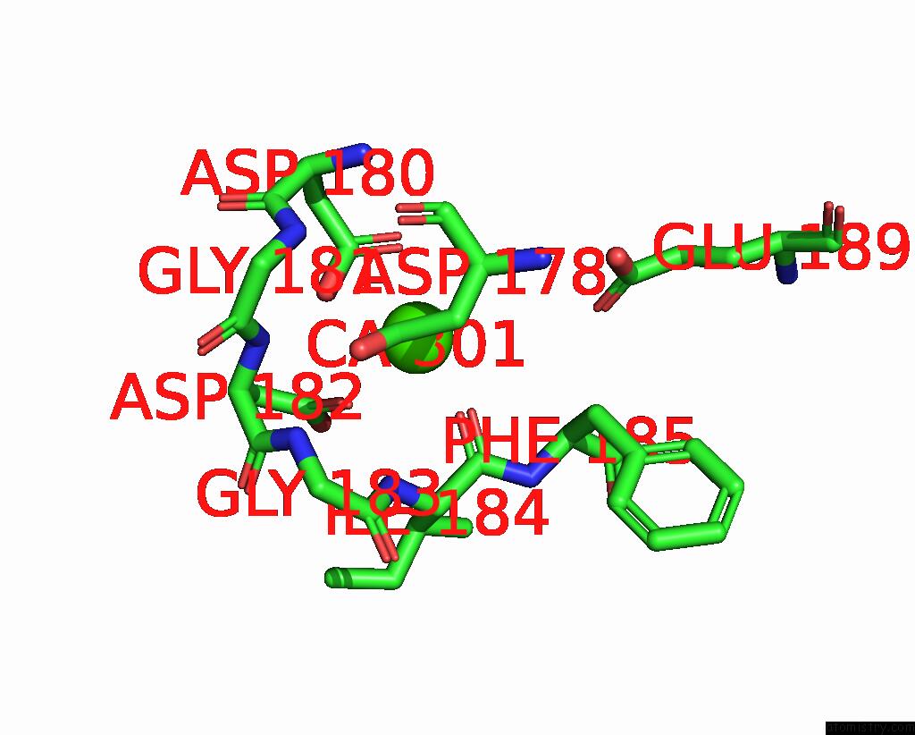
Mono view
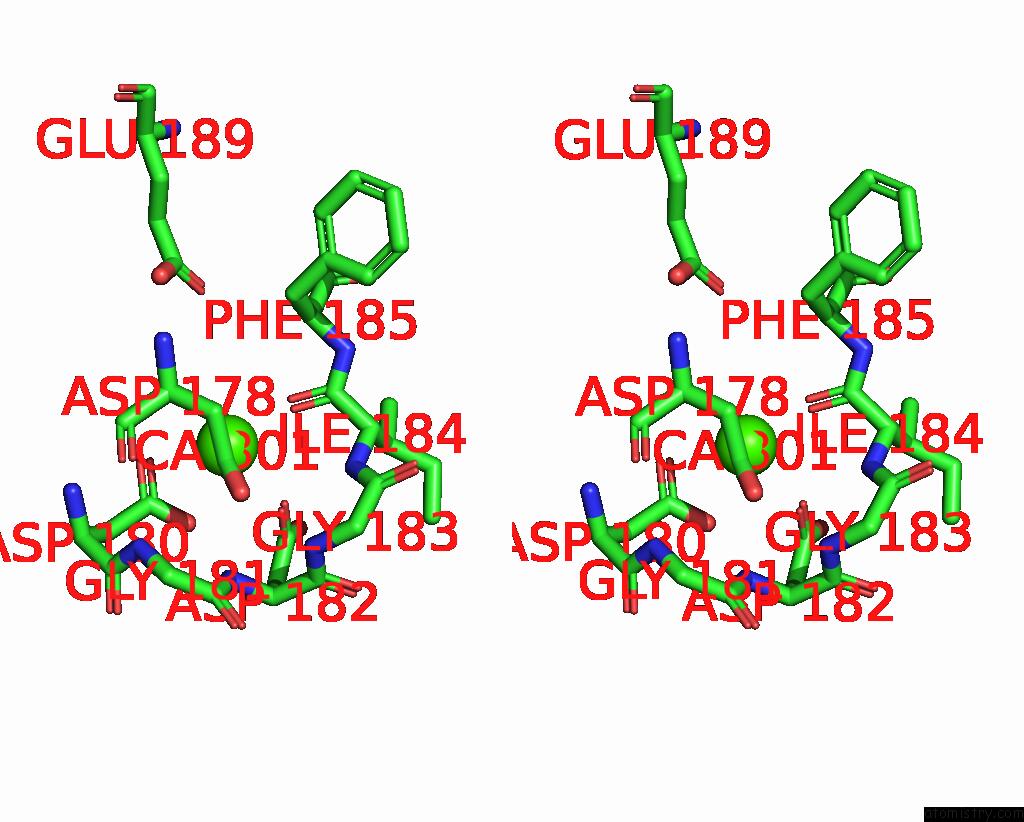
Stereo pair view
Mono view
Stereo pair view
A full contact list of Calcium with other atoms in the Ca binding
site number 1 of Crystal Structure of Ruminococcus Gnavus Immunoglobulin Binding Protein in Complex with 338E6 Fab within 5.0Å range:
|
Calcium binding site 2 out of 2 in 7kpj
Go back to
Calcium binding site 2 out
of 2 in the Crystal Structure of Ruminococcus Gnavus Immunoglobulin Binding Protein in Complex with 338E6 Fab
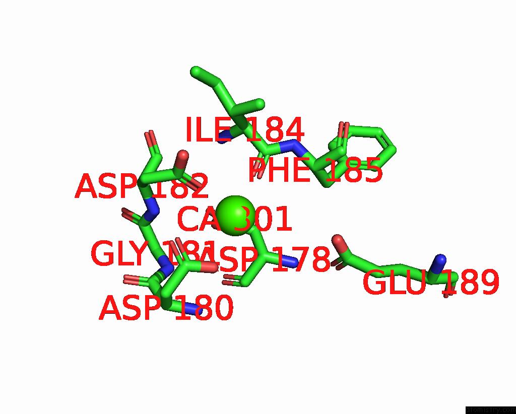
Mono view
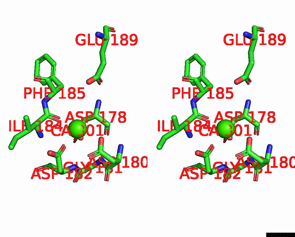
Stereo pair view
Mono view
Stereo pair view
A full contact list of Calcium with other atoms in the Ca binding
site number 2 of Crystal Structure of Ruminococcus Gnavus Immunoglobulin Binding Protein in Complex with 338E6 Fab within 5.0Å range:
|
Reference:
M.T.Borowska,
C.Drees,
A.E.Yarawsky,
M.Viswanathan,
S.M.Ryan,
J.J.Bunker,
A.B.Herr,
A.Bendelac,
E.J.Adams.
The Molecular Characterization of Antibody Binding to A Superantigen-Like Protein From A Commensal Microbe. Proc.Natl.Acad.Sci.Usa V. 118 2021.
ISSN: ESSN 1091-6490
PubMed: 34548394
DOI: 10.1073/PNAS.2023898118
Page generated: Wed Jul 9 22:59:32 2025
ISSN: ESSN 1091-6490
PubMed: 34548394
DOI: 10.1073/PNAS.2023898118
Last articles
Fe in 2YXOFe in 2YRS
Fe in 2YXC
Fe in 2YNM
Fe in 2YVJ
Fe in 2YP1
Fe in 2YU2
Fe in 2YU1
Fe in 2YQB
Fe in 2YOO