Calcium »
PDB 7kld-7l6g »
7kse »
Calcium in PDB 7kse: Crystal Structure of Prototype Foamy Virus Protease-Reverse Transcriptase Csh Mutant (Selenomethionine-Labeled)
Enzymatic activity of Crystal Structure of Prototype Foamy Virus Protease-Reverse Transcriptase Csh Mutant (Selenomethionine-Labeled)
All present enzymatic activity of Crystal Structure of Prototype Foamy Virus Protease-Reverse Transcriptase Csh Mutant (Selenomethionine-Labeled):
2.7.7.49; 2.7.7.7; 3.1.26.4;
2.7.7.49; 2.7.7.7; 3.1.26.4;
Protein crystallography data
The structure of Crystal Structure of Prototype Foamy Virus Protease-Reverse Transcriptase Csh Mutant (Selenomethionine-Labeled), PDB code: 7kse
was solved by
J.J.E.K.Harrison,
K.Das,
F.X.Ruiz,
E.Arnold,
with X-Ray Crystallography technique. A brief refinement statistics is given in the table below:
| Resolution Low / High (Å) | 50.00 / 3.00 |
| Space group | C 1 2 1 |
| Cell size a, b, c (Å), α, β, γ (°) | 240.375, 53.352, 74.922, 90, 100, 90 |
| R / Rfree (%) | 27.8 / 31.3 |
Calcium Binding Sites:
The binding sites of Calcium atom in the Crystal Structure of Prototype Foamy Virus Protease-Reverse Transcriptase Csh Mutant (Selenomethionine-Labeled)
(pdb code 7kse). This binding sites where shown within
5.0 Angstroms radius around Calcium atom.
In total only one binding site of Calcium was determined in the Crystal Structure of Prototype Foamy Virus Protease-Reverse Transcriptase Csh Mutant (Selenomethionine-Labeled), PDB code: 7kse:
In total only one binding site of Calcium was determined in the Crystal Structure of Prototype Foamy Virus Protease-Reverse Transcriptase Csh Mutant (Selenomethionine-Labeled), PDB code: 7kse:
Calcium binding site 1 out of 1 in 7kse
Go back to
Calcium binding site 1 out
of 1 in the Crystal Structure of Prototype Foamy Virus Protease-Reverse Transcriptase Csh Mutant (Selenomethionine-Labeled)
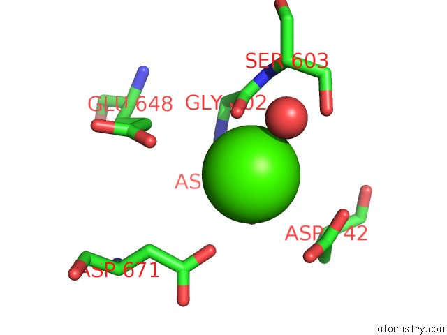
Mono view
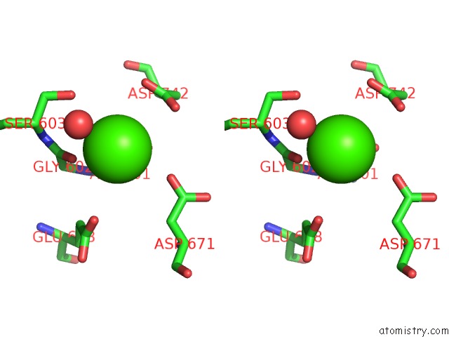
Stereo pair view
Mono view
Stereo pair view
A full contact list of Calcium with other atoms in the Ca binding
site number 1 of Crystal Structure of Prototype Foamy Virus Protease-Reverse Transcriptase Csh Mutant (Selenomethionine-Labeled) within 5.0Å range:
|
Reference:
J.J.E.K.Harrison,
S.Tuske,
K.Das,
F.X.Ruiz,
J.D.Bauman,
P.L.Boyer,
J.J.Destefano,
S.H.Hughes,
E.Arnold.
Crystal Structure of A Retroviral Polyprotein: Prototype Foamy Virus Protease-Reverse Transcriptase (Pr-Rt) Viruses V. 13 2021.
ISSN: ESSN 1999-4915
DOI: 10.3390/V13081495
Page generated: Wed Jul 9 23:00:15 2025
ISSN: ESSN 1999-4915
DOI: 10.3390/V13081495
Last articles
Fe in 2YXOFe in 2YRS
Fe in 2YXC
Fe in 2YNM
Fe in 2YVJ
Fe in 2YP1
Fe in 2YU2
Fe in 2YU1
Fe in 2YQB
Fe in 2YOO