Calcium »
PDB 7kld-7l6g »
7l29 »
Calcium in PDB 7l29: Crystal Structure of the Catalytic Domain of Human PDE3A Bound to Amp
Enzymatic activity of Crystal Structure of the Catalytic Domain of Human PDE3A Bound to Amp
All present enzymatic activity of Crystal Structure of the Catalytic Domain of Human PDE3A Bound to Amp:
3.1.4.17;
3.1.4.17;
Protein crystallography data
The structure of Crystal Structure of the Catalytic Domain of Human PDE3A Bound to Amp, PDB code: 7l29
was solved by
S.W.Horner,
C.Garvie,
with X-Ray Crystallography technique. A brief refinement statistics is given in the table below:
| Resolution Low / High (Å) | 47.90 / 2.08 |
| Space group | P 1 21 1 |
| Cell size a, b, c (Å), α, β, γ (°) | 82.415, 58.783, 156.996, 90, 90.74, 90 |
| R / Rfree (%) | 23.5 / 25.3 |
Other elements in 7l29:
The structure of Crystal Structure of the Catalytic Domain of Human PDE3A Bound to Amp also contains other interesting chemical elements:
| Magnesium | (Mg) | 4 atoms |
| Manganese | (Mn) | 4 atoms |
Calcium Binding Sites:
The binding sites of Calcium atom in the Crystal Structure of the Catalytic Domain of Human PDE3A Bound to Amp
(pdb code 7l29). This binding sites where shown within
5.0 Angstroms radius around Calcium atom.
In total only one binding site of Calcium was determined in the Crystal Structure of the Catalytic Domain of Human PDE3A Bound to Amp, PDB code: 7l29:
In total only one binding site of Calcium was determined in the Crystal Structure of the Catalytic Domain of Human PDE3A Bound to Amp, PDB code: 7l29:
Calcium binding site 1 out of 1 in 7l29
Go back to
Calcium binding site 1 out
of 1 in the Crystal Structure of the Catalytic Domain of Human PDE3A Bound to Amp
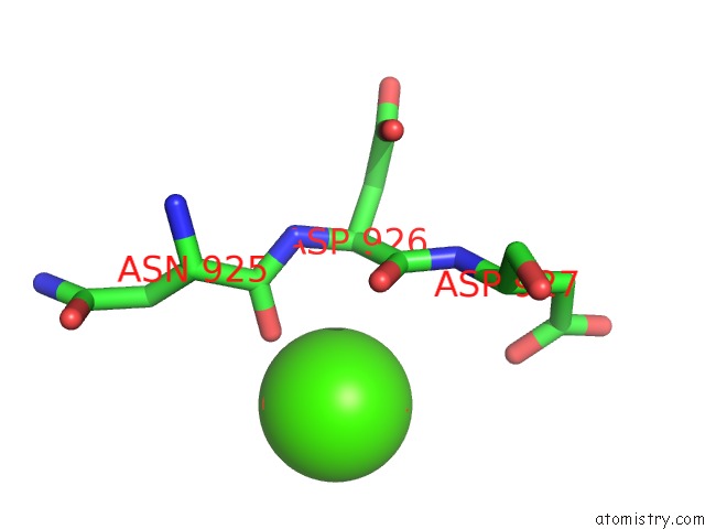
Mono view
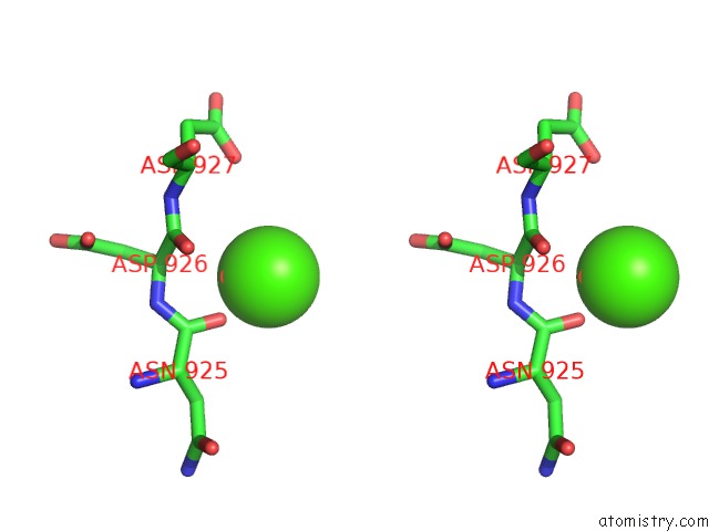
Stereo pair view
Mono view
Stereo pair view
A full contact list of Calcium with other atoms in the Ca binding
site number 1 of Crystal Structure of the Catalytic Domain of Human PDE3A Bound to Amp within 5.0Å range:
|
Reference:
C.W.Garvie,
S.W.Horner.
Structure of PDE3A-SLFN12 Complex Reveals Requirements For Activation of SLFN12 Rnase To Be Published.
Page generated: Wed Jul 9 23:02:19 2025
Last articles
Fe in 2YXOFe in 2YRS
Fe in 2YXC
Fe in 2YNM
Fe in 2YVJ
Fe in 2YP1
Fe in 2YU2
Fe in 2YU1
Fe in 2YQB
Fe in 2YOO