Calcium »
PDB 7kld-7l6g »
7l66 »
Calcium in PDB 7l66: C-Type Carbohydrate-Recognition Domain 4 of the Mannose Receptor Complexed with Methyl-Glcnac
Protein crystallography data
The structure of C-Type Carbohydrate-Recognition Domain 4 of the Mannose Receptor Complexed with Methyl-Glcnac, PDB code: 7l66
was solved by
W.I.Weis,
H.Feinberg,
with X-Ray Crystallography technique. A brief refinement statistics is given in the table below:
| Resolution Low / High (Å) | 34.44 / 1.75 |
| Space group | P 21 21 2 |
| Cell size a, b, c (Å), α, β, γ (°) | 54.484, 66.633, 34.436, 90, 90, 90 |
| R / Rfree (%) | 18.9 / 22 |
Calcium Binding Sites:
The binding sites of Calcium atom in the C-Type Carbohydrate-Recognition Domain 4 of the Mannose Receptor Complexed with Methyl-Glcnac
(pdb code 7l66). This binding sites where shown within
5.0 Angstroms radius around Calcium atom.
In total only one binding site of Calcium was determined in the C-Type Carbohydrate-Recognition Domain 4 of the Mannose Receptor Complexed with Methyl-Glcnac, PDB code: 7l66:
In total only one binding site of Calcium was determined in the C-Type Carbohydrate-Recognition Domain 4 of the Mannose Receptor Complexed with Methyl-Glcnac, PDB code: 7l66:
Calcium binding site 1 out of 1 in 7l66
Go back to
Calcium binding site 1 out
of 1 in the C-Type Carbohydrate-Recognition Domain 4 of the Mannose Receptor Complexed with Methyl-Glcnac
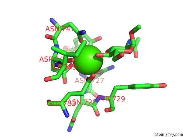
Mono view
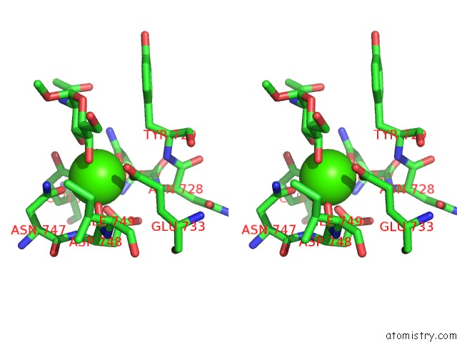
Stereo pair view
Mono view
Stereo pair view
A full contact list of Calcium with other atoms in the Ca binding
site number 1 of C-Type Carbohydrate-Recognition Domain 4 of the Mannose Receptor Complexed with Methyl-Glcnac within 5.0Å range:
|
Reference:
H.Feinberg,
S.A.F.Jegouzo,
Y.Lasanajak,
D.F.Smith,
K.Drickamer,
M.E.Taylor,
W.I.Weis.
Structural Analysis of Carbohydrate Binding By the Macrophage Mannose Receptor CD206 J.Biol.Chem. 2021.
ISSN: ESSN 1083-351X
Page generated: Wed Jul 9 23:03:55 2025
ISSN: ESSN 1083-351X
Last articles
Cl in 5JKACl in 5JKC
Cl in 5JK3
Cl in 5JJQ
Cl in 5JK0
Cl in 5JIR
Cl in 5JJX
Cl in 5JJV
Cl in 5JJM
Cl in 5JIZ