Calcium »
PDB 7l74-7lrx »
7l74 »
Calcium in PDB 7l74: Crystal Structure of Beta-Hexosyl Transferase From Hamamotoa (Sporobolomyces) Singularis Bound to Tris
Protein crystallography data
The structure of Crystal Structure of Beta-Hexosyl Transferase From Hamamotoa (Sporobolomyces) Singularis Bound to Tris, PDB code: 7l74
was solved by
S.F.Dagher,
B.F.P.Edwards,
F.Meilleur,
J.M.Bruno-Barcena,
with X-Ray Crystallography technique. A brief refinement statistics is given in the table below:
| Resolution Low / High (Å) | 29.96 / 2.25 |
| Space group | C 1 2 1 |
| Cell size a, b, c (Å), α, β, γ (°) | 196.319, 63.209, 105.006, 90, 100.42, 90 |
| R / Rfree (%) | 18.9 / 23.5 |
Calcium Binding Sites:
The binding sites of Calcium atom in the Crystal Structure of Beta-Hexosyl Transferase From Hamamotoa (Sporobolomyces) Singularis Bound to Tris
(pdb code 7l74). This binding sites where shown within
5.0 Angstroms radius around Calcium atom.
In total only one binding site of Calcium was determined in the Crystal Structure of Beta-Hexosyl Transferase From Hamamotoa (Sporobolomyces) Singularis Bound to Tris, PDB code: 7l74:
In total only one binding site of Calcium was determined in the Crystal Structure of Beta-Hexosyl Transferase From Hamamotoa (Sporobolomyces) Singularis Bound to Tris, PDB code: 7l74:
Calcium binding site 1 out of 1 in 7l74
Go back to
Calcium binding site 1 out
of 1 in the Crystal Structure of Beta-Hexosyl Transferase From Hamamotoa (Sporobolomyces) Singularis Bound to Tris
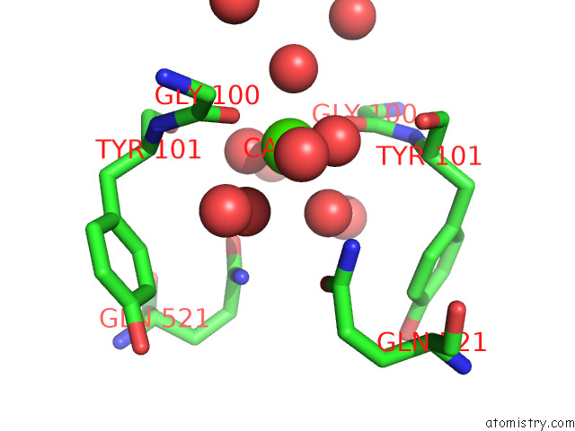
Mono view
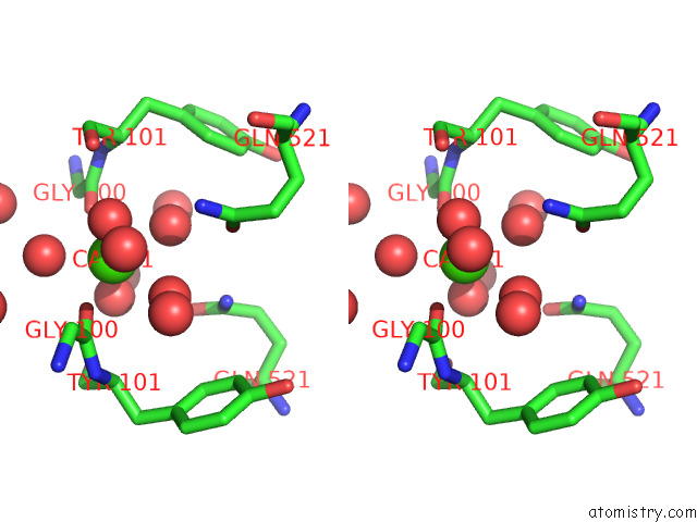
Stereo pair view
Mono view
Stereo pair view
A full contact list of Calcium with other atoms in the Ca binding
site number 1 of Crystal Structure of Beta-Hexosyl Transferase From Hamamotoa (Sporobolomyces) Singularis Bound to Tris within 5.0Å range:
|
Reference:
S.F.Dagher,
B.F.P.Edwards,
F.Meilleur,
J.M.Bruno-Barcena.
Structure and Mutagenic Analysis of the Beta-Hexosyltransferase From Hamamotoa (Sporobolomyces) Singularis To Be Published.
Page generated: Wed Jul 9 23:06:44 2025
Last articles
Fe in 2YXOFe in 2YRS
Fe in 2YXC
Fe in 2YNM
Fe in 2YVJ
Fe in 2YP1
Fe in 2YU2
Fe in 2YU1
Fe in 2YQB
Fe in 2YOO