Calcium »
PDB 7l74-7lrx »
7lri »
Calcium in PDB 7lri: Structure of Hiv-1 Reverse Transcriptase in Complex with Dna, Dttp, and Ca(2+) Ion
Enzymatic activity of Structure of Hiv-1 Reverse Transcriptase in Complex with Dna, Dttp, and Ca(2+) Ion
All present enzymatic activity of Structure of Hiv-1 Reverse Transcriptase in Complex with Dna, Dttp, and Ca(2+) Ion:
2.7.7.49; 2.7.7.7; 3.1.26.13;
2.7.7.49; 2.7.7.7; 3.1.26.13;
Protein crystallography data
The structure of Structure of Hiv-1 Reverse Transcriptase in Complex with Dna, Dttp, and Ca(2+) Ion, PDB code: 7lri
was solved by
A.Hoang,
F.X.Ruiz,
E.Arnold,
with X-Ray Crystallography technique. A brief refinement statistics is given in the table below:
| Resolution Low / High (Å) | 47.18 / 3.05 |
| Space group | P 1 21 1 |
| Cell size a, b, c (Å), α, β, γ (°) | 89.423, 131.073, 129.847, 90, 101.55, 90 |
| R / Rfree (%) | 21.1 / 24.4 |
Calcium Binding Sites:
The binding sites of Calcium atom in the Structure of Hiv-1 Reverse Transcriptase in Complex with Dna, Dttp, and Ca(2+) Ion
(pdb code 7lri). This binding sites where shown within
5.0 Angstroms radius around Calcium atom.
In total 2 binding sites of Calcium where determined in the Structure of Hiv-1 Reverse Transcriptase in Complex with Dna, Dttp, and Ca(2+) Ion, PDB code: 7lri:
Jump to Calcium binding site number: 1; 2;
In total 2 binding sites of Calcium where determined in the Structure of Hiv-1 Reverse Transcriptase in Complex with Dna, Dttp, and Ca(2+) Ion, PDB code: 7lri:
Jump to Calcium binding site number: 1; 2;
Calcium binding site 1 out of 2 in 7lri
Go back to
Calcium binding site 1 out
of 2 in the Structure of Hiv-1 Reverse Transcriptase in Complex with Dna, Dttp, and Ca(2+) Ion
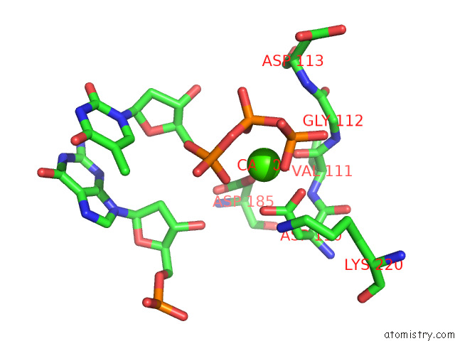
Mono view
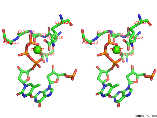
Stereo pair view
Mono view
Stereo pair view
A full contact list of Calcium with other atoms in the Ca binding
site number 1 of Structure of Hiv-1 Reverse Transcriptase in Complex with Dna, Dttp, and Ca(2+) Ion within 5.0Å range:
|
Calcium binding site 2 out of 2 in 7lri
Go back to
Calcium binding site 2 out
of 2 in the Structure of Hiv-1 Reverse Transcriptase in Complex with Dna, Dttp, and Ca(2+) Ion
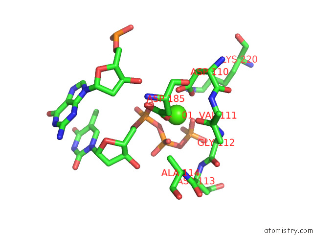
Mono view
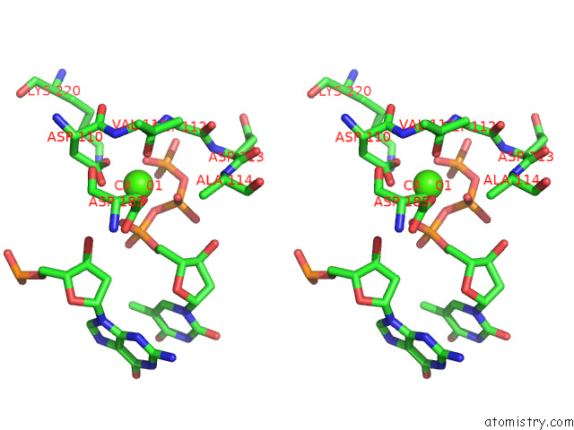
Stereo pair view
Mono view
Stereo pair view
A full contact list of Calcium with other atoms in the Ca binding
site number 2 of Structure of Hiv-1 Reverse Transcriptase in Complex with Dna, Dttp, and Ca(2+) Ion within 5.0Å range:
|
Reference:
F.X.Ruiz,
A.Hoang,
C.R.Dilmore,
J.J.Destefano,
E.Arnold.
Structural Basis of Hiv Inhibition By L-Nucleosides: Opportunities For Drug Development and Repurposing. Drug Discov Today V. 27 1832 2022.
ISSN: ESSN 1878-5832
PubMed: 35218925
DOI: 10.1016/J.DRUDIS.2022.02.016
Page generated: Wed Jul 9 23:16:50 2025
ISSN: ESSN 1878-5832
PubMed: 35218925
DOI: 10.1016/J.DRUDIS.2022.02.016
Last articles
Fe in 2YXOFe in 2YRS
Fe in 2YXC
Fe in 2YNM
Fe in 2YVJ
Fe in 2YP1
Fe in 2YU2
Fe in 2YU1
Fe in 2YQB
Fe in 2YOO