Calcium »
PDB 7lry-7m41 »
7lst »
Calcium in PDB 7lst: Ruminococcus Bromii AMY12-D392A with 63-A-D-Glucosyl-Maltotriosyl- Maltotriose
Enzymatic activity of Ruminococcus Bromii AMY12-D392A with 63-A-D-Glucosyl-Maltotriosyl- Maltotriose
All present enzymatic activity of Ruminococcus Bromii AMY12-D392A with 63-A-D-Glucosyl-Maltotriosyl- Maltotriose:
3.2.1.41;
3.2.1.41;
Protein crystallography data
The structure of Ruminococcus Bromii AMY12-D392A with 63-A-D-Glucosyl-Maltotriosyl- Maltotriose, PDB code: 7lst
was solved by
N.M.Koropatkin,
D.W.Cockburn,
H.A.Brown,
R.D.Kibler,
with X-Ray Crystallography technique. A brief refinement statistics is given in the table below:
| Resolution Low / High (Å) | 85.26 / 2.05 |
| Space group | P 21 21 21 |
| Cell size a, b, c (Å), α, β, γ (°) | 46.981, 99.107, 167.236, 90, 90, 90 |
| R / Rfree (%) | 18 / 23.2 |
Other elements in 7lst:
The structure of Ruminococcus Bromii AMY12-D392A with 63-A-D-Glucosyl-Maltotriosyl- Maltotriose also contains other interesting chemical elements:
| Sodium | (Na) | 1 atom |
Calcium Binding Sites:
The binding sites of Calcium atom in the Ruminococcus Bromii AMY12-D392A with 63-A-D-Glucosyl-Maltotriosyl- Maltotriose
(pdb code 7lst). This binding sites where shown within
5.0 Angstroms radius around Calcium atom.
In total only one binding site of Calcium was determined in the Ruminococcus Bromii AMY12-D392A with 63-A-D-Glucosyl-Maltotriosyl- Maltotriose, PDB code: 7lst:
In total only one binding site of Calcium was determined in the Ruminococcus Bromii AMY12-D392A with 63-A-D-Glucosyl-Maltotriosyl- Maltotriose, PDB code: 7lst:
Calcium binding site 1 out of 1 in 7lst
Go back to
Calcium binding site 1 out
of 1 in the Ruminococcus Bromii AMY12-D392A with 63-A-D-Glucosyl-Maltotriosyl- Maltotriose
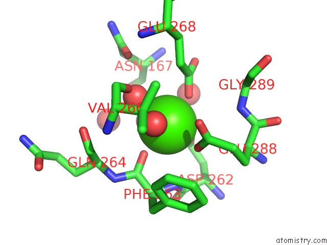
Mono view
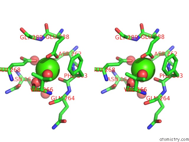
Stereo pair view
Mono view
Stereo pair view
A full contact list of Calcium with other atoms in the Ca binding
site number 1 of Ruminococcus Bromii AMY12-D392A with 63-A-D-Glucosyl-Maltotriosyl- Maltotriose within 5.0Å range:
|
Reference:
D.W.Cockburn,
R.Kibler,
H.A.Brown,
R.Duvall,
S.Morais,
E.Bayer,
N.M.Koropatkin.
Structure and Substrate Recognition By the Ruminococcus Bromii Amylosome Pullulanases. J.Struct.Biol. V. 213 07765 2021.
ISSN: ESSN 1095-8657
PubMed: 34186214
DOI: 10.1016/J.JSB.2021.107765
Page generated: Wed Jul 9 23:18:06 2025
ISSN: ESSN 1095-8657
PubMed: 34186214
DOI: 10.1016/J.JSB.2021.107765
Last articles
Fe in 2YXOFe in 2YRS
Fe in 2YXC
Fe in 2YNM
Fe in 2YVJ
Fe in 2YP1
Fe in 2YU2
Fe in 2YU1
Fe in 2YQB
Fe in 2YOO