Calcium »
PDB 7m43-7mg7 »
7mfl »
Calcium in PDB 7mfl: Structure of the Clostridium Perfringens GH89 in Complex with Beta- Hnjnac
Protein crystallography data
The structure of Structure of the Clostridium Perfringens GH89 in Complex with Beta- Hnjnac, PDB code: 7mfl
was solved by
A.B.Boraston,
with X-Ray Crystallography technique. A brief refinement statistics is given in the table below:
| Resolution Low / High (Å) | 29.79 / 2.00 |
| Space group | P 61 |
| Cell size a, b, c (Å), α, β, γ (°) | 91, 91, 252.812, 90, 90, 120 |
| R / Rfree (%) | 16.2 / 19.7 |
Calcium Binding Sites:
The binding sites of Calcium atom in the Structure of the Clostridium Perfringens GH89 in Complex with Beta- Hnjnac
(pdb code 7mfl). This binding sites where shown within
5.0 Angstroms radius around Calcium atom.
In total only one binding site of Calcium was determined in the Structure of the Clostridium Perfringens GH89 in Complex with Beta- Hnjnac, PDB code: 7mfl:
In total only one binding site of Calcium was determined in the Structure of the Clostridium Perfringens GH89 in Complex with Beta- Hnjnac, PDB code: 7mfl:
Calcium binding site 1 out of 1 in 7mfl
Go back to
Calcium binding site 1 out
of 1 in the Structure of the Clostridium Perfringens GH89 in Complex with Beta- Hnjnac
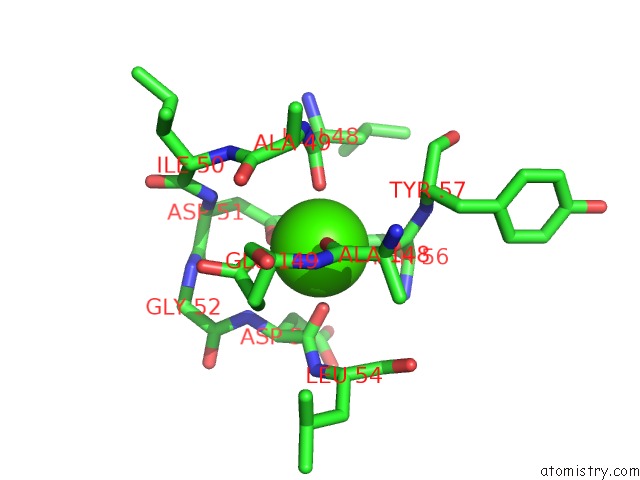
Mono view
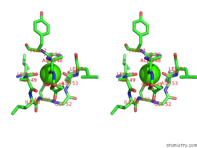
Stereo pair view
Mono view
Stereo pair view
A full contact list of Calcium with other atoms in the Ca binding
site number 1 of Structure of the Clostridium Perfringens GH89 in Complex with Beta- Hnjnac within 5.0Å range:
|
Reference:
S.Zhu,
Y.Jagadeesh,
A.T.Tran,
S.Imaeda,
A.Boraston,
D.S.Alonzi,
A.Poveda,
Y.Zhang,
J.Desire,
J.Charollais-Thoenig,
S.Demotz,
A.Kato,
T.D.Butters,
J.Jimenez-Barbero,
M.Sollogoub,
Y.Bleriot.
Iminosugar C-Glycosides Work As Pharmacological Chaperones of Naglu, A Glycosidase Involved in Mps Iiib Rare Disease*. Chemistry 2021.
ISSN: ISSN 0947-6539
PubMed: 34106504
DOI: 10.1002/CHEM.202101408
Page generated: Wed Jul 9 23:29:16 2025
ISSN: ISSN 0947-6539
PubMed: 34106504
DOI: 10.1002/CHEM.202101408
Last articles
Fe in 2YXOFe in 2YRS
Fe in 2YXC
Fe in 2YNM
Fe in 2YVJ
Fe in 2YP1
Fe in 2YU2
Fe in 2YU1
Fe in 2YQB
Fe in 2YOO