Calcium »
PDB 7nif-7o1w »
7nmx »
Calcium in PDB 7nmx: Crystal Structure of 14-3-3 Sigma in Complex with 13MER Amot-P130 Peptide and Fragment 12
Protein crystallography data
The structure of Crystal Structure of 14-3-3 Sigma in Complex with 13MER Amot-P130 Peptide and Fragment 12, PDB code: 7nmx
was solved by
F.Centorrino,
C.Ottmann,
with X-Ray Crystallography technique. A brief refinement statistics is given in the table below:
| Resolution Low / High (Å) | 29.89 / 2.30 |
| Space group | C 2 2 21 |
| Cell size a, b, c (Å), α, β, γ (°) | 82.469, 111.993, 62.699, 90, 90, 90 |
| R / Rfree (%) | 17.1 / 22.7 |
Other elements in 7nmx:
The structure of Crystal Structure of 14-3-3 Sigma in Complex with 13MER Amot-P130 Peptide and Fragment 12 also contains other interesting chemical elements:
| Chlorine | (Cl) | 3 atoms |
| Magnesium | (Mg) | 1 atom |
Calcium Binding Sites:
The binding sites of Calcium atom in the Crystal Structure of 14-3-3 Sigma in Complex with 13MER Amot-P130 Peptide and Fragment 12
(pdb code 7nmx). This binding sites where shown within
5.0 Angstroms radius around Calcium atom.
In total 2 binding sites of Calcium where determined in the Crystal Structure of 14-3-3 Sigma in Complex with 13MER Amot-P130 Peptide and Fragment 12, PDB code: 7nmx:
Jump to Calcium binding site number: 1; 2;
In total 2 binding sites of Calcium where determined in the Crystal Structure of 14-3-3 Sigma in Complex with 13MER Amot-P130 Peptide and Fragment 12, PDB code: 7nmx:
Jump to Calcium binding site number: 1; 2;
Calcium binding site 1 out of 2 in 7nmx
Go back to
Calcium binding site 1 out
of 2 in the Crystal Structure of 14-3-3 Sigma in Complex with 13MER Amot-P130 Peptide and Fragment 12
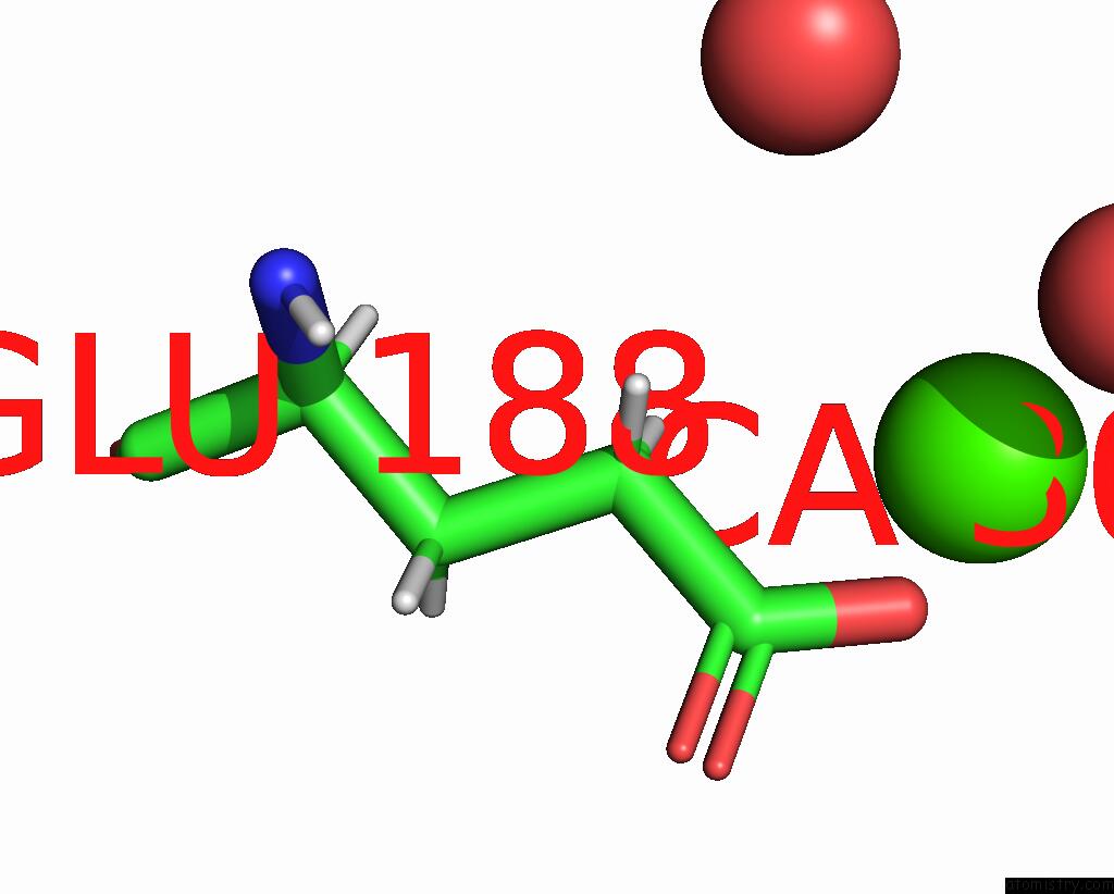
Mono view
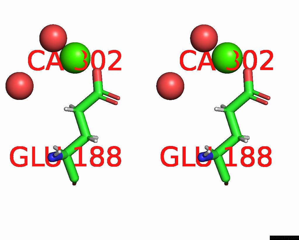
Stereo pair view
Mono view
Stereo pair view
A full contact list of Calcium with other atoms in the Ca binding
site number 1 of Crystal Structure of 14-3-3 Sigma in Complex with 13MER Amot-P130 Peptide and Fragment 12 within 5.0Å range:
|
Calcium binding site 2 out of 2 in 7nmx
Go back to
Calcium binding site 2 out
of 2 in the Crystal Structure of 14-3-3 Sigma in Complex with 13MER Amot-P130 Peptide and Fragment 12
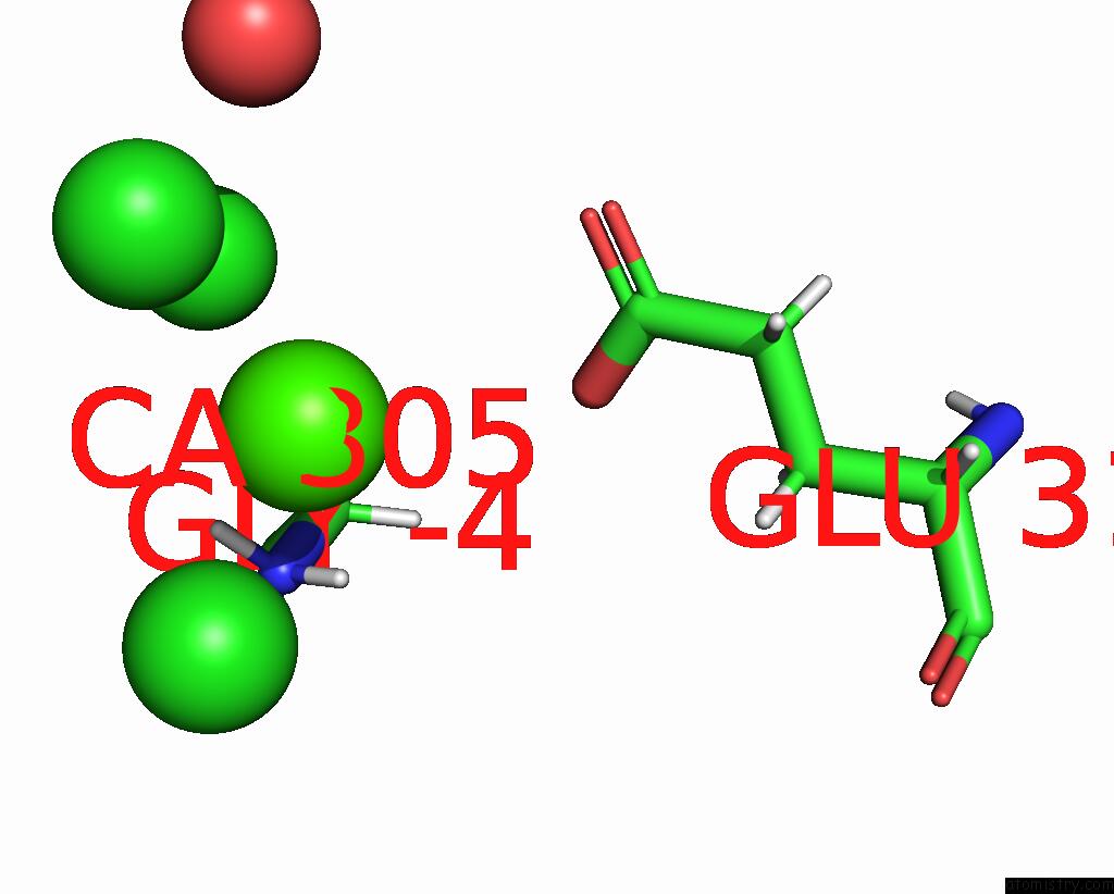
Mono view
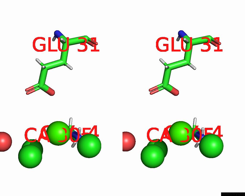
Stereo pair view
Mono view
Stereo pair view
A full contact list of Calcium with other atoms in the Ca binding
site number 2 of Crystal Structure of 14-3-3 Sigma in Complex with 13MER Amot-P130 Peptide and Fragment 12 within 5.0Å range:
|
Reference:
F.Centorrino,
B.Andlovic,
P.Cossar,
L.Brunsveld,
C.Ottmann.
Fragment-Based Exploration of the 14-3-3/Amot-P130 Interface. Curr Res Struct Biol V. 4 21 2022.
ISSN: ESSN 2665-928X
PubMed: 35036934
DOI: 10.1016/J.CRSTBI.2021.12.003
Page generated: Wed Jul 9 23:42:39 2025
ISSN: ESSN 2665-928X
PubMed: 35036934
DOI: 10.1016/J.CRSTBI.2021.12.003
Last articles
Cl in 5KC9Cl in 5KGE
Cl in 5KGA
Cl in 5KEZ
Cl in 5KDZ
Cl in 5KEG
Cl in 5KDY
Cl in 5KDT
Cl in 5KDB
Cl in 5KDQ