Calcium »
PDB 7nif-7o1w »
7nyj »
Calcium in PDB 7nyj: Structure of OBP1 From Varroa Destructor, Form P3<2>21
Protein crystallography data
The structure of Structure of OBP1 From Varroa Destructor, Form P3<2>21, PDB code: 7nyj
was solved by
C.Cambillau,
B.Amigues,
A.Roussel,
P.Leone,
A.Gaubert,
P.Pelosi,
with X-Ray Crystallography technique. A brief refinement statistics is given in the table below:
| Resolution Low / High (Å) | 46.59 / 1.81 |
| Space group | P 32 2 1 |
| Cell size a, b, c (Å), α, β, γ (°) | 53.8, 53.8, 91.67, 90, 90, 120 |
| R / Rfree (%) | 18.6 / 22.6 |
Other elements in 7nyj:
The structure of Structure of OBP1 From Varroa Destructor, Form P3<2>21 also contains other interesting chemical elements:
| Sodium | (Na) | 1 atom |
Calcium Binding Sites:
The binding sites of Calcium atom in the Structure of OBP1 From Varroa Destructor, Form P3<2>21
(pdb code 7nyj). This binding sites where shown within
5.0 Angstroms radius around Calcium atom.
In total 2 binding sites of Calcium where determined in the Structure of OBP1 From Varroa Destructor, Form P3<2>21, PDB code: 7nyj:
Jump to Calcium binding site number: 1; 2;
In total 2 binding sites of Calcium where determined in the Structure of OBP1 From Varroa Destructor, Form P3<2>21, PDB code: 7nyj:
Jump to Calcium binding site number: 1; 2;
Calcium binding site 1 out of 2 in 7nyj
Go back to
Calcium binding site 1 out
of 2 in the Structure of OBP1 From Varroa Destructor, Form P3<2>21
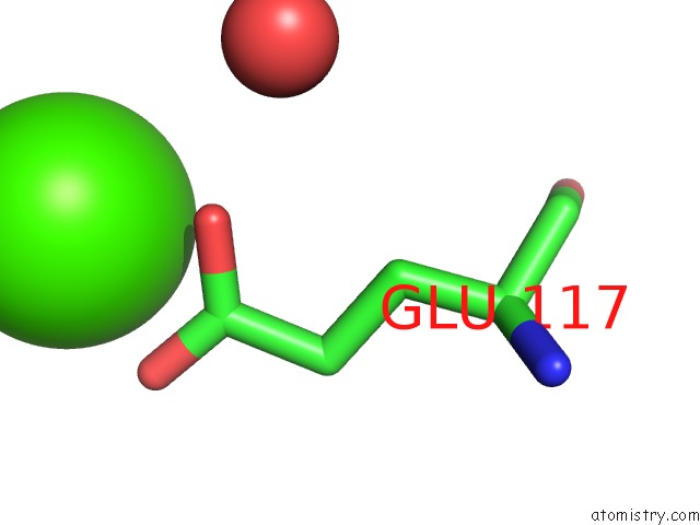
Mono view
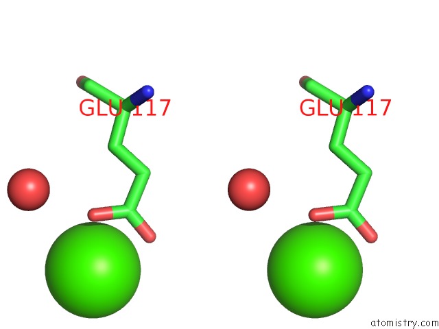
Stereo pair view
Mono view
Stereo pair view
A full contact list of Calcium with other atoms in the Ca binding
site number 1 of Structure of OBP1 From Varroa Destructor, Form P3<2>21 within 5.0Å range:
|
Calcium binding site 2 out of 2 in 7nyj
Go back to
Calcium binding site 2 out
of 2 in the Structure of OBP1 From Varroa Destructor, Form P3<2>21
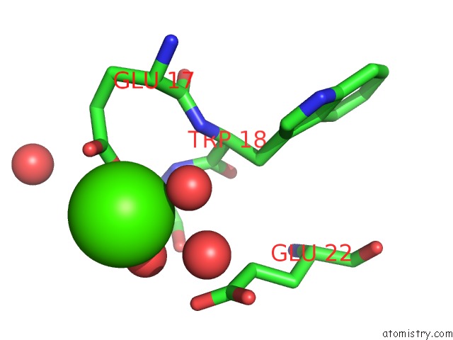
Mono view
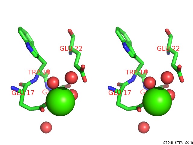
Stereo pair view
Mono view
Stereo pair view
A full contact list of Calcium with other atoms in the Ca binding
site number 2 of Structure of OBP1 From Varroa Destructor, Form P3<2>21 within 5.0Å range:
|
Reference:
B.Amigues,
J.Zhu,
A.Gaubert,
S.Arena,
G.Renzone,
P.Leone,
I.M.Fischer,
H.Paulsen,
W.Knoll,
A.Scaloni,
A.Roussel,
C.Cambillau,
P.Pelosi.
A New Non-Classical Fold of Varroa Odorant-Binding Proteins Reveals A Wide Open Internal Cavity. Sci Rep V. 11 13172 2021.
ISSN: ESSN 2045-2322
PubMed: 34162975
DOI: 10.1038/S41598-021-92604-2
Page generated: Wed Jul 9 23:46:26 2025
ISSN: ESSN 2045-2322
PubMed: 34162975
DOI: 10.1038/S41598-021-92604-2
Last articles
Fe in 2YXOFe in 2YRS
Fe in 2YXC
Fe in 2YNM
Fe in 2YVJ
Fe in 2YP1
Fe in 2YU2
Fe in 2YU1
Fe in 2YQB
Fe in 2YOO