Calcium »
PDB 7o1y-7or1 »
7o20 »
Calcium in PDB 7o20: X-Ray Structure of Furin in Complex with the Guanylhydrazone-Based Inhibitor 3 (MI300)
Enzymatic activity of X-Ray Structure of Furin in Complex with the Guanylhydrazone-Based Inhibitor 3 (MI300)
All present enzymatic activity of X-Ray Structure of Furin in Complex with the Guanylhydrazone-Based Inhibitor 3 (MI300):
3.4.21.75;
3.4.21.75;
Protein crystallography data
The structure of X-Ray Structure of Furin in Complex with the Guanylhydrazone-Based Inhibitor 3 (MI300), PDB code: 7o20
was solved by
S.O.Dahms,
H.Brandstetter,
with X-Ray Crystallography technique. A brief refinement statistics is given in the table below:
| Resolution Low / High (Å) | 45.72 / 1.80 |
| Space group | P 65 2 2 |
| Cell size a, b, c (Å), α, β, γ (°) | 130.316, 130.316, 156.037, 90, 90, 120 |
| R / Rfree (%) | 16.2 / 17.6 |
Other elements in 7o20:
The structure of X-Ray Structure of Furin in Complex with the Guanylhydrazone-Based Inhibitor 3 (MI300) also contains other interesting chemical elements:
| Chlorine | (Cl) | 1 atom |
| Sodium | (Na) | 2 atoms |
Calcium Binding Sites:
The binding sites of Calcium atom in the X-Ray Structure of Furin in Complex with the Guanylhydrazone-Based Inhibitor 3 (MI300)
(pdb code 7o20). This binding sites where shown within
5.0 Angstroms radius around Calcium atom.
In total 3 binding sites of Calcium where determined in the X-Ray Structure of Furin in Complex with the Guanylhydrazone-Based Inhibitor 3 (MI300), PDB code: 7o20:
Jump to Calcium binding site number: 1; 2; 3;
In total 3 binding sites of Calcium where determined in the X-Ray Structure of Furin in Complex with the Guanylhydrazone-Based Inhibitor 3 (MI300), PDB code: 7o20:
Jump to Calcium binding site number: 1; 2; 3;
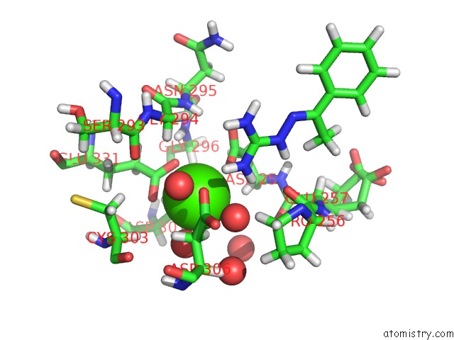
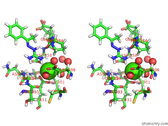
Calcium binding site 1 out of 3 in 7o20
Go back to
Calcium binding site 1 out
of 3 in the X-Ray Structure of Furin in Complex with the Guanylhydrazone-Based Inhibitor 3 (MI300)
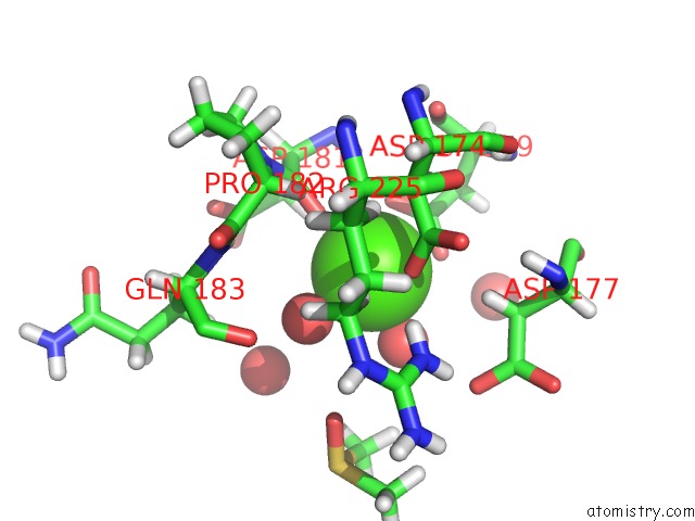
Mono view
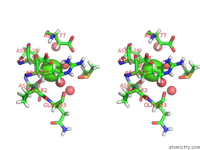
Stereo pair view
Mono view
Stereo pair view
A full contact list of Calcium with other atoms in the Ca binding
site number 1 of X-Ray Structure of Furin in Complex with the Guanylhydrazone-Based Inhibitor 3 (MI300) within 5.0Å range:
|
Calcium binding site 2 out of 3 in 7o20
Go back to
Calcium binding site 2 out
of 3 in the X-Ray Structure of Furin in Complex with the Guanylhydrazone-Based Inhibitor 3 (MI300)
Mono view
Stereo pair view
Mono view
Stereo pair view
A full contact list of Calcium with other atoms in the Ca binding
site number 2 of X-Ray Structure of Furin in Complex with the Guanylhydrazone-Based Inhibitor 3 (MI300) within 5.0Å range:
|
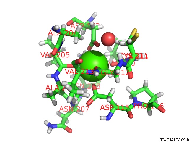
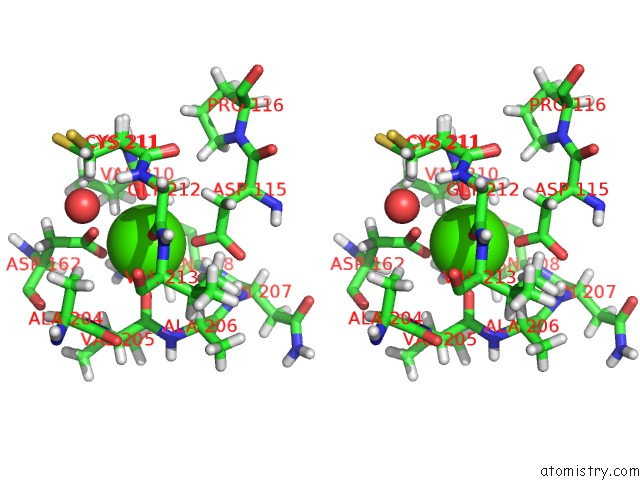
Calcium binding site 3 out of 3 in 7o20
Go back to
Calcium binding site 3 out
of 3 in the X-Ray Structure of Furin in Complex with the Guanylhydrazone-Based Inhibitor 3 (MI300)
Mono view
Stereo pair view
Mono view
Stereo pair view
A full contact list of Calcium with other atoms in the Ca binding
site number 3 of X-Ray Structure of Furin in Complex with the Guanylhydrazone-Based Inhibitor 3 (MI300) within 5.0Å range:
|
Reference:
S.O.Dahms,
T.Haider,
G.Klebe,
T.Steinmetzer,
H.Brandstetter.
Off-State-Specific Inhibition of the Proprotein Convertase Furin. Acs Chem.Biol. 2021.
ISSN: ESSN 1554-8937
PubMed: 34415722
DOI: 10.1021/ACSCHEMBIO.1C00411
Page generated: Wed Jul 9 23:47:55 2025
ISSN: ESSN 1554-8937
PubMed: 34415722
DOI: 10.1021/ACSCHEMBIO.1C00411
Last articles
Fe in 2YXOFe in 2YRS
Fe in 2YXC
Fe in 2YNM
Fe in 2YVJ
Fe in 2YP1
Fe in 2YU2
Fe in 2YU1
Fe in 2YQB
Fe in 2YOO