Calcium »
PDB 7o1y-7or1 »
7ob5 »
Calcium in PDB 7ob5: Crystal Structure of 14-3-3 Sigma in Complex with LDB1 Phosphopeptide
Protein crystallography data
The structure of Crystal Structure of 14-3-3 Sigma in Complex with LDB1 Phosphopeptide, PDB code: 7ob5
was solved by
F.Centorrino,
B.Andlovic,
C.Ottmann,
with X-Ray Crystallography technique. A brief refinement statistics is given in the table below:
| Resolution Low / High (Å) | 41.30 / 1.80 |
| Space group | C 2 2 21 |
| Cell size a, b, c (Å), α, β, γ (°) | 82.59, 112.188, 62.573, 90, 90, 90 |
| R / Rfree (%) | 13.8 / 17.1 |
Other elements in 7ob5:
The structure of Crystal Structure of 14-3-3 Sigma in Complex with LDB1 Phosphopeptide also contains other interesting chemical elements:
| Magnesium | (Mg) | 1 atom |
| Chlorine | (Cl) | 1 atom |
Calcium Binding Sites:
The binding sites of Calcium atom in the Crystal Structure of 14-3-3 Sigma in Complex with LDB1 Phosphopeptide
(pdb code 7ob5). This binding sites where shown within
5.0 Angstroms radius around Calcium atom.
In total 2 binding sites of Calcium where determined in the Crystal Structure of 14-3-3 Sigma in Complex with LDB1 Phosphopeptide, PDB code: 7ob5:
Jump to Calcium binding site number: 1; 2;
In total 2 binding sites of Calcium where determined in the Crystal Structure of 14-3-3 Sigma in Complex with LDB1 Phosphopeptide, PDB code: 7ob5:
Jump to Calcium binding site number: 1; 2;
Calcium binding site 1 out of 2 in 7ob5
Go back to
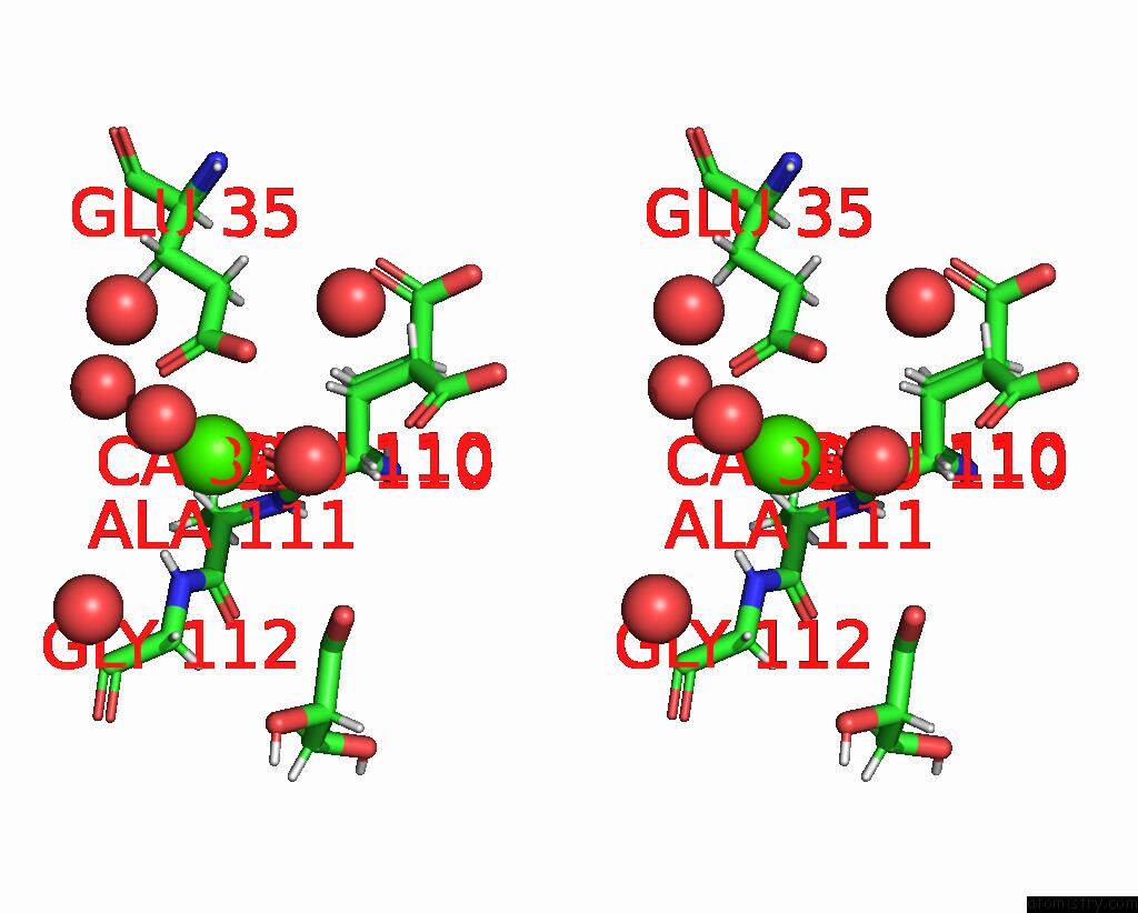
Calcium binding site 1 out
of 2 in the Crystal Structure of 14-3-3 Sigma in Complex with LDB1 Phosphopeptide

Mono view

Stereo pair view
Mono view
Stereo pair view
A full contact list of Calcium with other atoms in the Ca binding
site number 1 of Crystal Structure of 14-3-3 Sigma in Complex with LDB1 Phosphopeptide within 5.0Å range:
|
Calcium binding site 2 out of 2 in 7ob5
Go back to
Calcium binding site 2 out
of 2 in the Crystal Structure of 14-3-3 Sigma in Complex with LDB1 Phosphopeptide

Mono view
Stereo pair view
Mono view
Stereo pair view
A full contact list of Calcium with other atoms in the Ca binding
site number 2 of Crystal Structure of 14-3-3 Sigma in Complex with LDB1 Phosphopeptide within 5.0Å range:
|
Reference:
B.Andlovic,
G.Heilmann,
S.Ninck,
S.Andrei,
F.Centorrino,
Y.Higuchi,
N.Kato,
L.Brunsveld,
S.Menninger,
A.Choidas,
A.Wolf,
M.Kaiser,
J.Eickhoff,
C.Ottmann.
Inf Alpha Primes Ovarian Cancer Cells For Fusicoccin-Induced Cell Death Via Stabilization of 14-3-3 Protein-Protein Interactions To Be Published.
Page generated: Wed Jul 9 23:50:00 2025
Last articles
F in 7NVVF in 7NVO
F in 7NTH
F in 7NTI
F in 7NPC
F in 7NRG
F in 7NR5
F in 7NQS
F in 7NOS
F in 7NP5