Calcium »
PDB 7or8-7p9r »
7ozq »
Calcium in PDB 7ozq: Crystal Structure of Archaeal L7AE Bound to Eukaryotic Kink-Loop
Protein crystallography data
The structure of Crystal Structure of Archaeal L7AE Bound to Eukaryotic Kink-Loop, PDB code: 7ozq
was solved by
S.Hoefler,
P.Lukat,
T.Carlomagno,
W.Blankenfeldt,
with X-Ray Crystallography technique. A brief refinement statistics is given in the table below:
| Resolution Low / High (Å) | 54.50 / 1.91 |
| Space group | C 1 2 1 |
| Cell size a, b, c (Å), α, β, γ (°) | 109.391, 61.598, 138.611, 90, 108.36, 90 |
| R / Rfree (%) | 17.3 / 21.9 |
Other elements in 7ozq:
The structure of Crystal Structure of Archaeal L7AE Bound to Eukaryotic Kink-Loop also contains other interesting chemical elements:
| Chlorine | (Cl) | 1 atom |
| Sodium | (Na) | 6 atoms |
Calcium Binding Sites:
The binding sites of Calcium atom in the Crystal Structure of Archaeal L7AE Bound to Eukaryotic Kink-Loop
(pdb code 7ozq). This binding sites where shown within
5.0 Angstroms radius around Calcium atom.
In total 3 binding sites of Calcium where determined in the Crystal Structure of Archaeal L7AE Bound to Eukaryotic Kink-Loop, PDB code: 7ozq:
Jump to Calcium binding site number: 1; 2; 3;
In total 3 binding sites of Calcium where determined in the Crystal Structure of Archaeal L7AE Bound to Eukaryotic Kink-Loop, PDB code: 7ozq:
Jump to Calcium binding site number: 1; 2; 3;
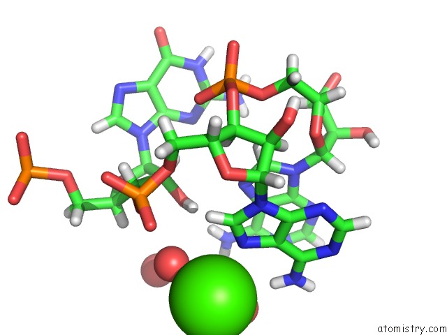
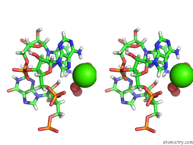
Calcium binding site 1 out of 3 in 7ozq
Go back to
Calcium binding site 1 out
of 3 in the Crystal Structure of Archaeal L7AE Bound to Eukaryotic Kink-Loop
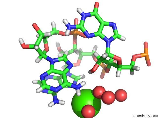
Mono view
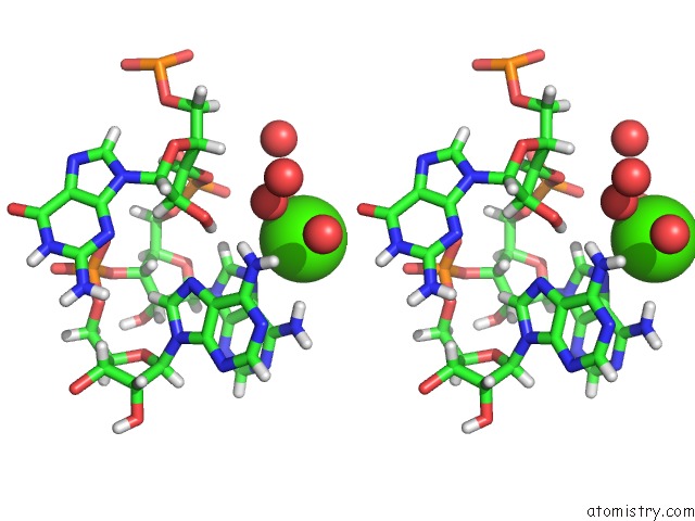
Stereo pair view
Mono view
Stereo pair view
A full contact list of Calcium with other atoms in the Ca binding
site number 1 of Crystal Structure of Archaeal L7AE Bound to Eukaryotic Kink-Loop within 5.0Å range:
|
Calcium binding site 2 out of 3 in 7ozq
Go back to
Calcium binding site 2 out
of 3 in the Crystal Structure of Archaeal L7AE Bound to Eukaryotic Kink-Loop
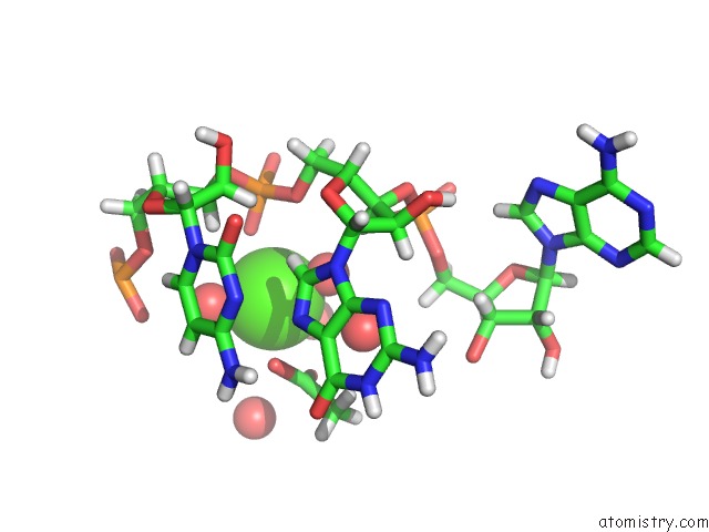
Mono view
Stereo pair view
Mono view
Stereo pair view
A full contact list of Calcium with other atoms in the Ca binding
site number 2 of Crystal Structure of Archaeal L7AE Bound to Eukaryotic Kink-Loop within 5.0Å range:
|
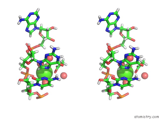
Calcium binding site 3 out of 3 in 7ozq
Go back to
Calcium binding site 3 out
of 3 in the Crystal Structure of Archaeal L7AE Bound to Eukaryotic Kink-Loop
Mono view
Stereo pair view
Mono view
Stereo pair view
A full contact list of Calcium with other atoms in the Ca binding
site number 3 of Crystal Structure of Archaeal L7AE Bound to Eukaryotic Kink-Loop within 5.0Å range:
|
Reference:
S.Hofler,
P.Lukat,
W.Blankenfeldt,
T.Carlomagno.
Eukaryotic Box C/D Methylation Machinery Has Two Non-Symmetric Protein Assembly Sites. Sci Rep V. 11 17561 2021.
ISSN: ESSN 2045-2322
PubMed: 34475498
DOI: 10.1038/S41598-021-97030-Y
Page generated: Wed Jul 9 23:58:10 2025
ISSN: ESSN 2045-2322
PubMed: 34475498
DOI: 10.1038/S41598-021-97030-Y
Last articles
Fe in 2YXOFe in 2YRS
Fe in 2YXC
Fe in 2YNM
Fe in 2YVJ
Fe in 2YP1
Fe in 2YU2
Fe in 2YU1
Fe in 2YQB
Fe in 2YOO