Calcium »
PDB 7or8-7p9r »
7p01 »
Calcium in PDB 7p01: Structure of the Maltase BAAG2 From Blastobotrys Adeninivorans in Complex with Acarbose
Enzymatic activity of Structure of the Maltase BAAG2 From Blastobotrys Adeninivorans in Complex with Acarbose
All present enzymatic activity of Structure of the Maltase BAAG2 From Blastobotrys Adeninivorans in Complex with Acarbose:
3.2.1.20;
3.2.1.20;
Protein crystallography data
The structure of Structure of the Maltase BAAG2 From Blastobotrys Adeninivorans in Complex with Acarbose, PDB code: 7p01
was solved by
K.Ernits,
T.Visnapuu,
K.Persson,
with X-Ray Crystallography technique. A brief refinement statistics is given in the table below:
| Resolution Low / High (Å) | 46.32 / 2.12 |
| Space group | P 1 21 1 |
| Cell size a, b, c (Å), α, β, γ (°) | 68.03, 78.08, 121.93, 90, 94.1, 90 |
| R / Rfree (%) | 18.4 / 19.5 |
Calcium Binding Sites:
The binding sites of Calcium atom in the Structure of the Maltase BAAG2 From Blastobotrys Adeninivorans in Complex with Acarbose
(pdb code 7p01). This binding sites where shown within
5.0 Angstroms radius around Calcium atom.
In total 2 binding sites of Calcium where determined in the Structure of the Maltase BAAG2 From Blastobotrys Adeninivorans in Complex with Acarbose, PDB code: 7p01:
Jump to Calcium binding site number: 1; 2;
In total 2 binding sites of Calcium where determined in the Structure of the Maltase BAAG2 From Blastobotrys Adeninivorans in Complex with Acarbose, PDB code: 7p01:
Jump to Calcium binding site number: 1; 2;
Calcium binding site 1 out of 2 in 7p01
Go back to
Calcium binding site 1 out
of 2 in the Structure of the Maltase BAAG2 From Blastobotrys Adeninivorans in Complex with Acarbose
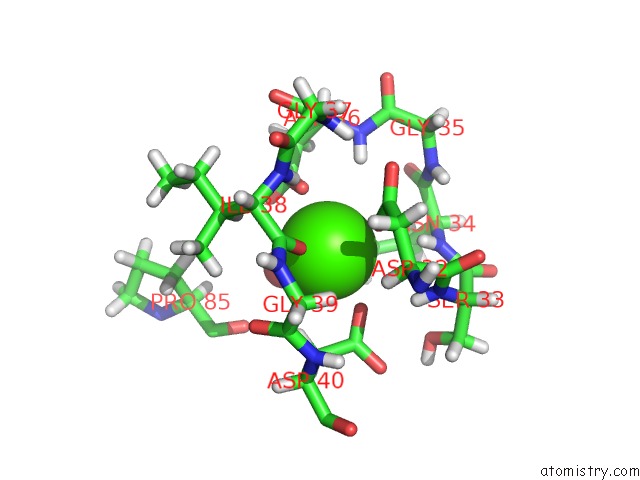
Mono view

Stereo pair view
Mono view
Stereo pair view
A full contact list of Calcium with other atoms in the Ca binding
site number 1 of Structure of the Maltase BAAG2 From Blastobotrys Adeninivorans in Complex with Acarbose within 5.0Å range:
|
Calcium binding site 2 out of 2 in 7p01
Go back to
Calcium binding site 2 out
of 2 in the Structure of the Maltase BAAG2 From Blastobotrys Adeninivorans in Complex with Acarbose
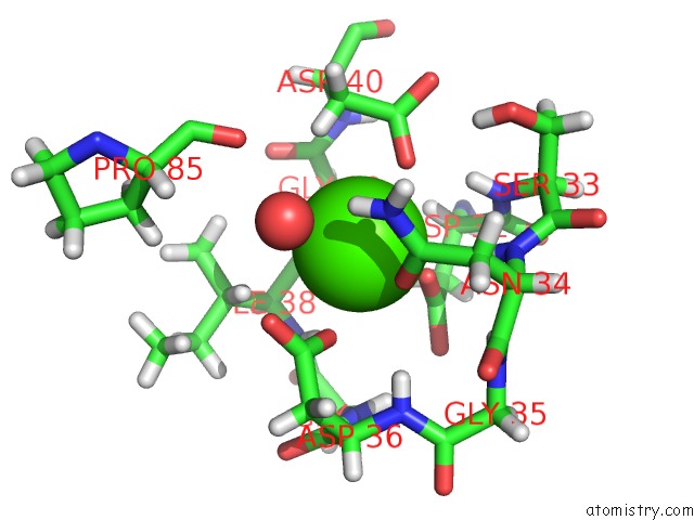
Mono view
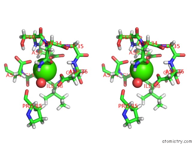
Stereo pair view
Mono view
Stereo pair view
A full contact list of Calcium with other atoms in the Ca binding
site number 2 of Structure of the Maltase BAAG2 From Blastobotrys Adeninivorans in Complex with Acarbose within 5.0Å range:
|
Reference:
K.Ernits,
C.Kjeldsen,
K.Persson,
E.Grigor,
T.Alamae,
T.Visnapuu.
Structural Insight Into A Yeast Maltase-the BAAG2 From Blastobotrys Adeninivorans with Transglycosylating Activity J Fungi V. 7 2021.
DOI: /10.3390/JOF7100816
Page generated: Wed Jul 9 23:58:38 2025
DOI: /10.3390/JOF7100816
Last articles
Cl in 8D7DCl in 8D7J
Cl in 8D7C
Cl in 8D7B
Cl in 8D77
Cl in 8D75
Cl in 8D7A
Cl in 8D6I
Cl in 8D6B
Cl in 8D69