Calcium in PDB 7p70: The Pdz-Domain of SNTB1 Complexed with the Pdz-Binding Motif of HPV35- E6
Protein crystallography data
The structure of The Pdz-Domain of SNTB1 Complexed with the Pdz-Binding Motif of HPV35- E6, PDB code: 7p70
was solved by
G.Gogl,
A.Cousido-Siah,
G.Trave,
with X-Ray Crystallography technique. A brief refinement statistics is given in the table below:
Resolution Low / High (Å)
|
45.90 /
2.00
|
Space group
|
P 21 21 21
|
Cell size a, b, c (Å), α, β, γ (°)
|
54.8,
60.58,
140.64,
90,
90,
90
|
R / Rfree (%)
|
18.3 /
20.9
|
Calcium Binding Sites:
The binding sites of Calcium atom in the The Pdz-Domain of SNTB1 Complexed with the Pdz-Binding Motif of HPV35- E6
(pdb code 7p70). This binding sites where shown within
5.0 Angstroms radius around Calcium atom.
In total 5 binding sites of Calcium where determined in the
The Pdz-Domain of SNTB1 Complexed with the Pdz-Binding Motif of HPV35- E6, PDB code: 7p70:
Jump to Calcium binding site number:
1;
2;
3;
4;
5;
Calcium binding site 1 out
of 5 in 7p70
Go back to
Calcium Binding Sites List in 7p70
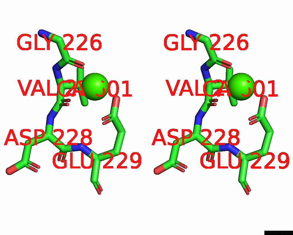
Calcium binding site 1 out
of 5 in the The Pdz-Domain of SNTB1 Complexed with the Pdz-Binding Motif of HPV35- E6
Mono view
Stereo pair view
|
A full contact list of Calcium with other atoms in the Ca binding
site number 1 of The Pdz-Domain of SNTB1 Complexed with the Pdz-Binding Motif of HPV35- E6 within 5.0Å range:
probe
|
atom
|
residue
|
distance (Å)
|
B
|
Occ
|
A:Ca601
b:73.4
occ:1.00
|
O
|
A:GLY226
|
2.5
|
50.4
|
1.0
|
OE1
|
A:GLU229
|
2.6
|
57.5
|
1.0
|
O
|
A:VAL227
|
2.7
|
48.9
|
1.0
|
OE2
|
A:GLU229
|
2.9
|
56.8
|
1.0
|
CD
|
A:GLU229
|
3.1
|
49.9
|
1.0
|
C
|
A:GLY226
|
3.5
|
43.6
|
1.0
|
C
|
A:VAL227
|
3.9
|
43.0
|
1.0
|
CA
|
A:GLY226
|
4.3
|
45.9
|
1.0
|
N
|
A:VAL227
|
4.3
|
44.0
|
1.0
|
CA
|
A:VAL227
|
4.5
|
50.5
|
1.0
|
CG
|
A:GLU229
|
4.6
|
39.1
|
1.0
|
CB
|
A:VAL227
|
4.7
|
40.4
|
1.0
|
N
|
A:GLU229
|
4.9
|
39.0
|
1.0
|
N
|
A:ASP228
|
5.0
|
43.6
|
1.0
|
|
Calcium binding site 2 out
of 5 in 7p70
Go back to
Calcium Binding Sites List in 7p70
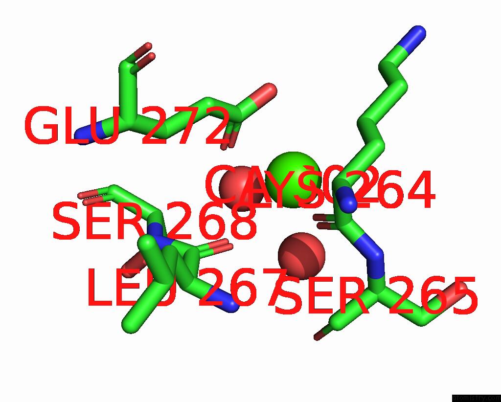
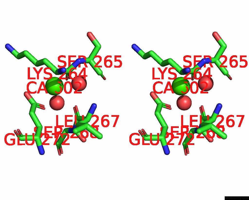
Calcium binding site 2 out
of 5 in the The Pdz-Domain of SNTB1 Complexed with the Pdz-Binding Motif of HPV35- E6
Mono view
Stereo pair view
|
A full contact list of Calcium with other atoms in the Ca binding
site number 2 of The Pdz-Domain of SNTB1 Complexed with the Pdz-Binding Motif of HPV35- E6 within 5.0Å range:
probe
|
atom
|
residue
|
distance (Å)
|
B
|
Occ
|
A:Ca602
b:82.8
occ:1.00
|
O
|
A:HOH783
|
2.4
|
59.4
|
1.0
|
O
|
A:LYS264
|
2.4
|
48.2
|
1.0
|
OE1
|
A:GLU272
|
2.4
|
65.5
|
1.0
|
O
|
A:HOH731
|
2.5
|
73.0
|
1.0
|
CD
|
A:GLU272
|
3.0
|
58.5
|
1.0
|
O
|
A:LEU267
|
3.1
|
37.2
|
1.0
|
OE2
|
A:GLU272
|
3.1
|
66.1
|
1.0
|
C
|
A:LYS264
|
3.5
|
43.1
|
1.0
|
CG
|
A:LYS264
|
3.6
|
56.2
|
1.0
|
CA
|
A:LYS264
|
4.2
|
43.9
|
1.0
|
C
|
A:LEU267
|
4.2
|
37.5
|
1.0
|
CG
|
A:GLU272
|
4.3
|
42.2
|
1.0
|
CB
|
A:LYS264
|
4.5
|
46.2
|
1.0
|
N
|
A:SER265
|
4.5
|
36.4
|
1.0
|
CA
|
A:SER268
|
4.6
|
30.9
|
1.0
|
CE
|
A:LYS264
|
4.6
|
67.8
|
1.0
|
CD
|
A:LYS264
|
4.7
|
63.4
|
1.0
|
CA
|
A:SER265
|
4.7
|
39.3
|
1.0
|
N
|
A:SER268
|
4.9
|
32.4
|
1.0
|
CB
|
A:GLU272
|
5.0
|
31.9
|
1.0
|
|
Calcium binding site 3 out
of 5 in 7p70
Go back to
Calcium Binding Sites List in 7p70
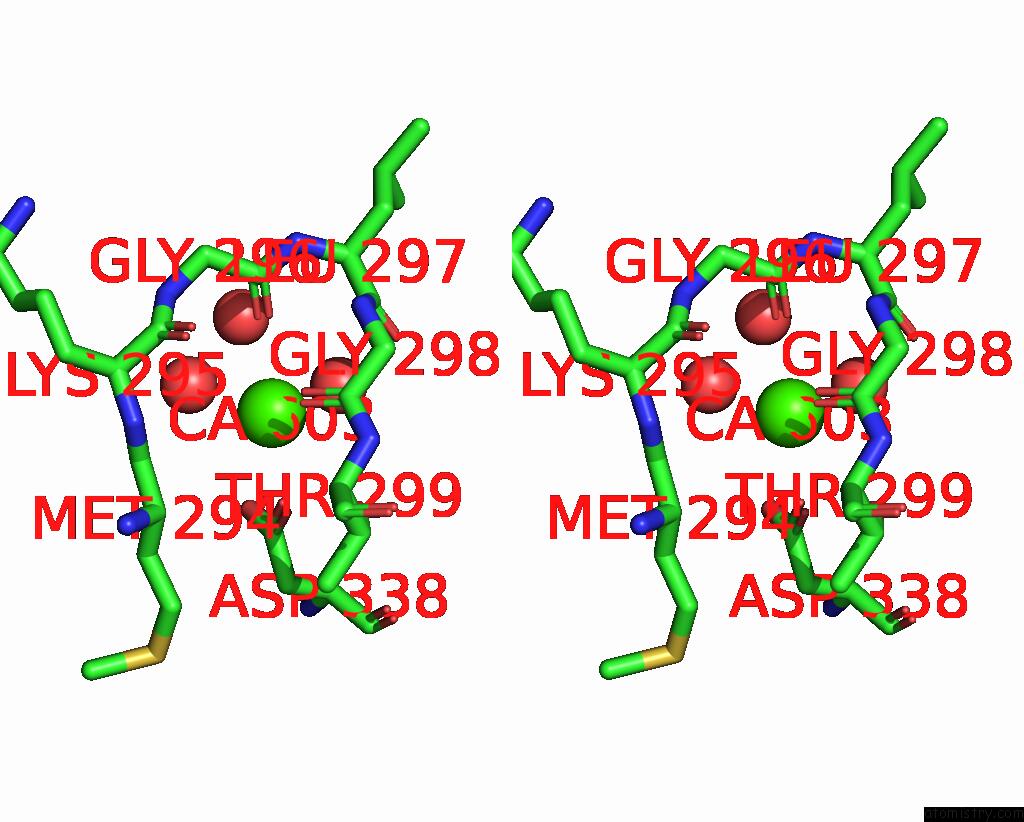
Calcium binding site 3 out
of 5 in the The Pdz-Domain of SNTB1 Complexed with the Pdz-Binding Motif of HPV35- E6
Mono view
Stereo pair view
|
A full contact list of Calcium with other atoms in the Ca binding
site number 3 of The Pdz-Domain of SNTB1 Complexed with the Pdz-Binding Motif of HPV35- E6 within 5.0Å range:
probe
|
atom
|
residue
|
distance (Å)
|
B
|
Occ
|
A:Ca603
b:80.0
occ:1.00
|
O
|
A:HOH824
|
2.5
|
51.3
|
1.0
|
OD1
|
A:ASP338
|
2.5
|
70.5
|
1.0
|
O
|
A:HOH873
|
2.5
|
59.0
|
1.0
|
O
|
A:GLY296
|
2.6
|
53.5
|
1.0
|
O
|
A:HOH821
|
2.8
|
57.6
|
1.0
|
O
|
A:GLY298
|
3.0
|
61.8
|
1.0
|
OD2
|
A:ASP338
|
3.2
|
75.9
|
1.0
|
CG
|
A:ASP338
|
3.2
|
68.8
|
1.0
|
C
|
A:GLY296
|
3.7
|
55.2
|
1.0
|
O
|
A:MET294
|
3.8
|
49.5
|
1.0
|
OG1
|
A:THR299
|
3.9
|
54.0
|
1.0
|
O
|
A:HOH852
|
3.9
|
52.5
|
1.0
|
O
|
A:LEU297
|
3.9
|
62.6
|
1.0
|
C
|
A:MET294
|
3.9
|
44.5
|
1.0
|
C
|
A:GLY298
|
3.9
|
59.9
|
1.0
|
C
|
A:LEU297
|
4.1
|
55.9
|
1.0
|
C
|
A:LYS295
|
4.2
|
57.4
|
1.0
|
N
|
A:LYS295
|
4.2
|
48.0
|
1.0
|
O
|
A:LYS295
|
4.3
|
64.6
|
1.0
|
N
|
A:GLY296
|
4.3
|
53.0
|
1.0
|
CA
|
A:MET294
|
4.4
|
38.2
|
1.0
|
N
|
A:GLY298
|
4.4
|
56.4
|
1.0
|
CA
|
A:LYS295
|
4.6
|
58.2
|
1.0
|
N
|
A:LEU297
|
4.6
|
54.4
|
1.0
|
CA
|
A:LEU297
|
4.6
|
46.5
|
1.0
|
CA
|
A:GLY296
|
4.6
|
52.6
|
1.0
|
CB
|
A:ASP338
|
4.7
|
63.9
|
1.0
|
CA
|
A:GLY298
|
4.7
|
60.1
|
1.0
|
N
|
A:THR299
|
4.8
|
57.0
|
1.0
|
CA
|
A:THR299
|
4.9
|
48.5
|
1.0
|
CB
|
A:THR299
|
5.0
|
53.2
|
1.0
|
|
Calcium binding site 4 out
of 5 in 7p70
Go back to
Calcium Binding Sites List in 7p70
Calcium binding site 4 out
of 5 in the The Pdz-Domain of SNTB1 Complexed with the Pdz-Binding Motif of HPV35- E6
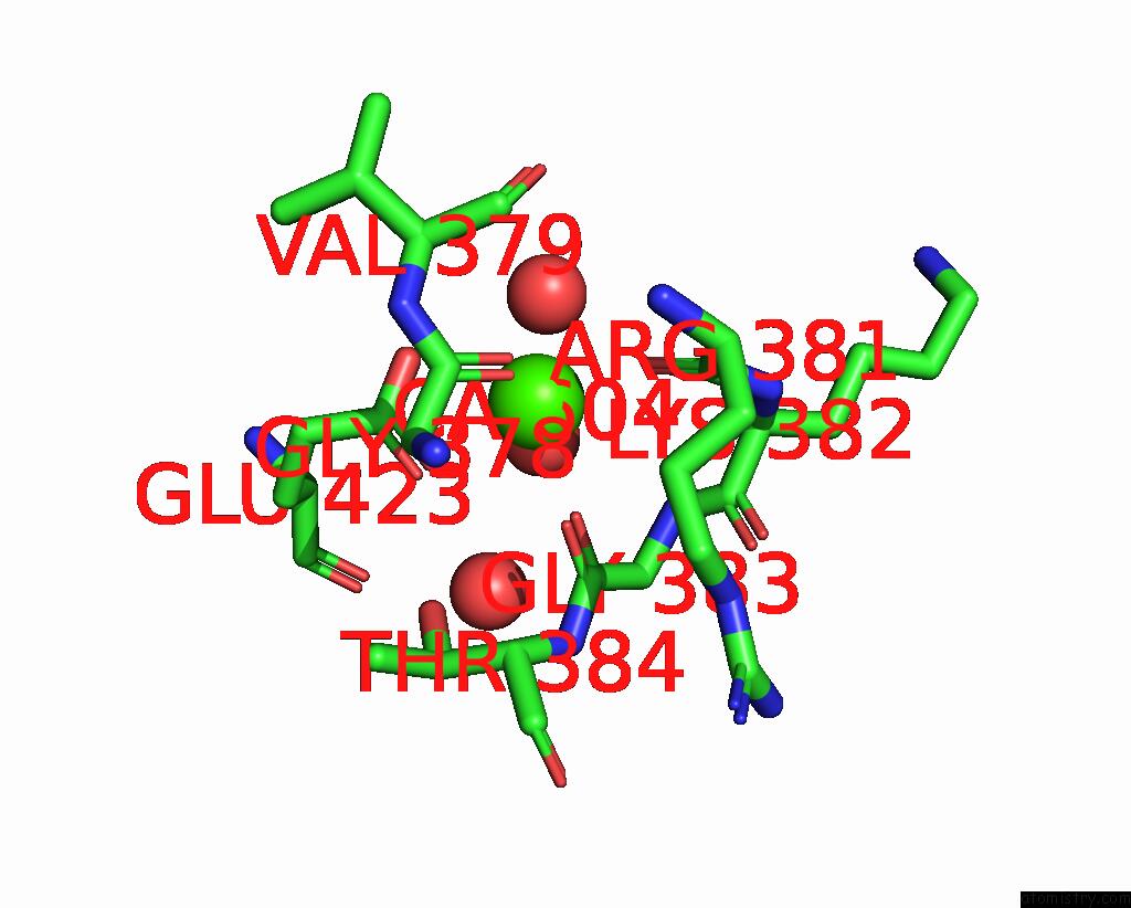
Mono view
Stereo pair view
|
A full contact list of Calcium with other atoms in the Ca binding
site number 4 of The Pdz-Domain of SNTB1 Complexed with the Pdz-Binding Motif of HPV35- E6 within 5.0Å range:
probe
|
atom
|
residue
|
distance (Å)
|
B
|
Occ
|
A:Ca604
b:49.1
occ:1.00
|
O
|
A:ARG381
|
2.4
|
47.1
|
1.0
|
O
|
A:GLY378
|
2.4
|
41.6
|
1.0
|
O
|
A:GLY383
|
2.4
|
43.0
|
1.0
|
O
|
A:HOH879
|
2.4
|
57.9
|
1.0
|
OE1
|
A:GLU423
|
2.5
|
42.6
|
1.0
|
OE2
|
A:GLU423
|
2.6
|
40.8
|
1.0
|
O
|
A:HOH912
|
2.8
|
53.8
|
1.0
|
CD
|
A:GLU423
|
2.9
|
44.0
|
1.0
|
C
|
A:GLY378
|
3.3
|
43.1
|
1.0
|
C
|
A:GLY383
|
3.5
|
43.5
|
1.0
|
C
|
A:ARG381
|
3.6
|
52.2
|
1.0
|
N
|
A:GLY383
|
3.6
|
47.0
|
1.0
|
CA
|
A:GLY383
|
4.0
|
49.7
|
1.0
|
C
|
A:LYS382
|
4.1
|
50.6
|
1.0
|
CA
|
A:GLY378
|
4.1
|
44.5
|
1.0
|
N
|
A:VAL379
|
4.2
|
39.1
|
1.0
|
CA
|
A:VAL379
|
4.3
|
41.4
|
1.0
|
CA
|
A:LYS382
|
4.3
|
53.8
|
1.0
|
N
|
A:LYS382
|
4.4
|
50.9
|
1.0
|
N
|
A:ARG381
|
4.4
|
50.2
|
1.0
|
CG
|
A:GLU423
|
4.4
|
36.7
|
1.0
|
C
|
A:VAL379
|
4.5
|
43.8
|
1.0
|
OG1
|
A:THR384
|
4.5
|
44.7
|
1.0
|
N
|
A:THR384
|
4.6
|
47.4
|
1.0
|
O
|
A:VAL379
|
4.6
|
50.1
|
1.0
|
CA
|
A:ARG381
|
4.6
|
52.1
|
1.0
|
O
|
A:HOH723
|
4.6
|
45.2
|
1.0
|
CG
|
A:ARG381
|
4.6
|
45.6
|
1.0
|
O
|
A:LYS382
|
4.9
|
53.6
|
1.0
|
CA
|
A:THR384
|
4.9
|
48.2
|
1.0
|
|
Calcium binding site 5 out
of 5 in 7p70
Go back to
Calcium Binding Sites List in 7p70
Calcium binding site 5 out
of 5 in the The Pdz-Domain of SNTB1 Complexed with the Pdz-Binding Motif of HPV35- E6
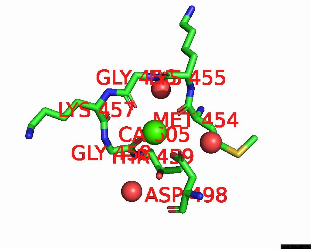
Mono view
Stereo pair view
|
A full contact list of Calcium with other atoms in the Ca binding
site number 5 of The Pdz-Domain of SNTB1 Complexed with the Pdz-Binding Motif of HPV35- E6 within 5.0Å range:
probe
|
atom
|
residue
|
distance (Å)
|
B
|
Occ
|
A:Ca605
b:29.0
occ:1.00
|
O
|
A:MET454
|
2.3
|
27.3
|
1.0
|
O
|
A:GLY456
|
2.4
|
29.4
|
1.0
|
O
|
A:GLY458
|
2.4
|
29.0
|
1.0
|
OD2
|
A:ASP498
|
2.5
|
29.0
|
1.0
|
OD1
|
A:ASP498
|
2.5
|
28.8
|
1.0
|
CG
|
A:ASP498
|
2.8
|
28.4
|
1.0
|
C
|
A:MET454
|
3.4
|
21.3
|
1.0
|
C
|
A:GLY458
|
3.6
|
35.3
|
1.0
|
C
|
A:GLY456
|
3.6
|
28.1
|
1.0
|
CA
|
A:MET454
|
4.0
|
25.4
|
1.0
|
N
|
A:GLY458
|
4.0
|
33.3
|
1.0
|
N
|
A:GLY456
|
4.1
|
26.7
|
1.0
|
CA
|
A:GLY458
|
4.3
|
29.4
|
1.0
|
C
|
A:LYS457
|
4.3
|
31.9
|
1.0
|
C
|
A:LYS455
|
4.3
|
27.1
|
1.0
|
CB
|
A:ASP498
|
4.3
|
26.7
|
1.0
|
OG1
|
A:THR459
|
4.4
|
32.1
|
1.0
|
N
|
A:LYS455
|
4.5
|
24.7
|
1.0
|
CA
|
A:GLY456
|
4.5
|
26.2
|
1.0
|
O
|
A:LYS455
|
4.5
|
29.1
|
1.0
|
CB
|
A:MET454
|
4.6
|
25.4
|
1.0
|
N
|
A:THR459
|
4.6
|
26.8
|
1.0
|
N
|
A:LYS457
|
4.6
|
30.2
|
1.0
|
O
|
A:HOH729
|
4.6
|
25.1
|
1.0
|
O
|
A:HOH894
|
4.7
|
52.8
|
1.0
|
CA
|
A:LYS457
|
4.7
|
28.9
|
1.0
|
O
|
A:LYS457
|
4.7
|
30.4
|
1.0
|
CA
|
A:LYS455
|
4.8
|
27.5
|
1.0
|
CA
|
A:THR459
|
4.8
|
30.3
|
1.0
|
O
|
A:HOH742
|
5.0
|
39.7
|
1.0
|
|
Reference:
G.Gogl,
A.Cousido-Siah,
G.Trave.
The Pdz-Domain of SNTB1 Complexed with the Pdz-Binding Motif of HPV35-E6 To Be Published.
Page generated: Fri Jul 19 02:44:46 2024
|