Calcium in PDB 7p71: The Pdz Domain of MAGI1_2 Complexed with the Pdz-Binding Motif of HPV35-E6
Protein crystallography data
The structure of The Pdz Domain of MAGI1_2 Complexed with the Pdz-Binding Motif of HPV35-E6, PDB code: 7p71
was solved by
G.Gogl,
A.Cousido-Siah,
G.Trave,
with X-Ray Crystallography technique. A brief refinement statistics is given in the table below:
Resolution Low / High (Å)
|
47.64 /
2.60
|
Space group
|
C 1 2 1
|
Cell size a, b, c (Å), α, β, γ (°)
|
192.13,
61.03,
98.99,
90,
97.33,
90
|
R / Rfree (%)
|
24.1 /
28.1
|
Calcium Binding Sites:
The binding sites of Calcium atom in the The Pdz Domain of MAGI1_2 Complexed with the Pdz-Binding Motif of HPV35-E6
(pdb code 7p71). This binding sites where shown within
5.0 Angstroms radius around Calcium atom.
In total 8 binding sites of Calcium where determined in the
The Pdz Domain of MAGI1_2 Complexed with the Pdz-Binding Motif of HPV35-E6, PDB code: 7p71:
Jump to Calcium binding site number:
1;
2;
3;
4;
5;
6;
7;
8;
Calcium binding site 1 out
of 8 in 7p71
Go back to
Calcium Binding Sites List in 7p71
Calcium binding site 1 out
of 8 in the The Pdz Domain of MAGI1_2 Complexed with the Pdz-Binding Motif of HPV35-E6
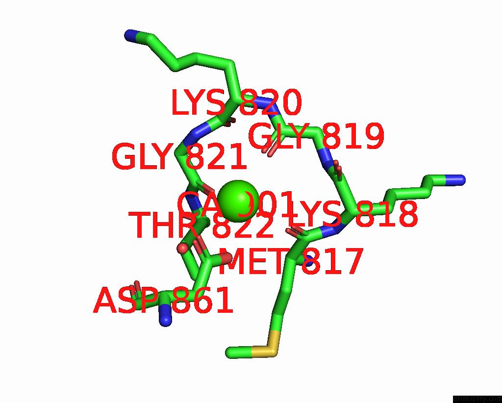
Mono view
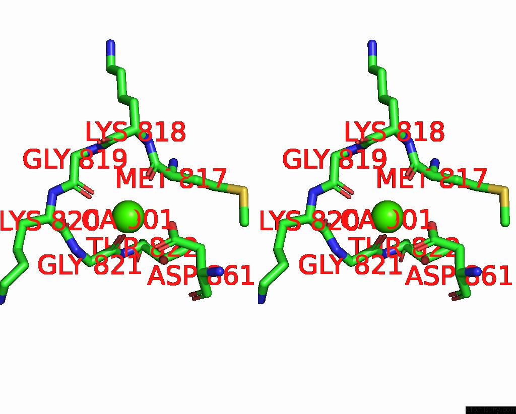
Stereo pair view
|
A full contact list of Calcium with other atoms in the Ca binding
site number 1 of The Pdz Domain of MAGI1_2 Complexed with the Pdz-Binding Motif of HPV35-E6 within 5.0Å range:
probe
|
atom
|
residue
|
distance (Å)
|
B
|
Occ
|
A:Ca901
b:125.0
occ:1.00
|
O
|
A:GLY819
|
2.4
|
60.8
|
1.0
|
O
|
A:GLY821
|
2.5
|
82.9
|
1.0
|
OD1
|
A:ASP861
|
2.5
|
118.1
|
1.0
|
O
|
A:MET817
|
2.5
|
89.3
|
1.0
|
OD2
|
A:ASP861
|
2.6
|
111.7
|
1.0
|
CG
|
A:ASP861
|
2.9
|
107.8
|
1.0
|
C
|
A:MET817
|
3.5
|
76.5
|
1.0
|
C
|
A:GLY821
|
3.6
|
75.9
|
1.0
|
C
|
A:GLY819
|
3.7
|
60.0
|
1.0
|
N
|
A:GLY821
|
3.9
|
66.3
|
1.0
|
CA
|
A:MET817
|
3.9
|
71.9
|
1.0
|
OG1
|
A:THR822
|
4.2
|
77.8
|
1.0
|
C
|
A:LYS820
|
4.2
|
68.6
|
1.0
|
CA
|
A:GLY821
|
4.3
|
70.0
|
1.0
|
N
|
A:GLY819
|
4.3
|
66.7
|
1.0
|
CB
|
A:ASP861
|
4.4
|
94.9
|
1.0
|
C
|
A:LYS818
|
4.4
|
75.1
|
1.0
|
N
|
A:LYS818
|
4.5
|
73.8
|
1.0
|
CA
|
A:LYS820
|
4.5
|
65.6
|
1.0
|
CB
|
A:MET817
|
4.5
|
65.5
|
1.0
|
N
|
A:LYS820
|
4.6
|
61.6
|
1.0
|
O
|
A:LYS818
|
4.6
|
81.7
|
1.0
|
CA
|
A:GLY819
|
4.6
|
61.1
|
1.0
|
N
|
A:THR822
|
4.7
|
76.3
|
1.0
|
O
|
A:LYS820
|
4.8
|
74.4
|
1.0
|
CA
|
A:LYS818
|
4.9
|
74.3
|
1.0
|
CA
|
A:THR822
|
4.9
|
73.0
|
1.0
|
|
Calcium binding site 2 out
of 8 in 7p71
Go back to
Calcium Binding Sites List in 7p71
Calcium binding site 2 out
of 8 in the The Pdz Domain of MAGI1_2 Complexed with the Pdz-Binding Motif of HPV35-E6
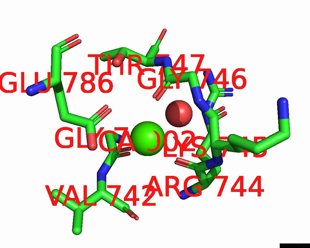
Mono view
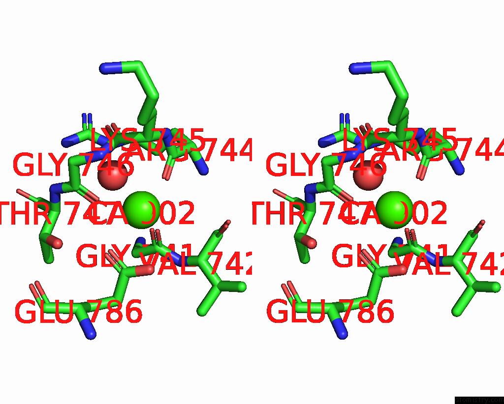
Stereo pair view
|
A full contact list of Calcium with other atoms in the Ca binding
site number 2 of The Pdz Domain of MAGI1_2 Complexed with the Pdz-Binding Motif of HPV35-E6 within 5.0Å range:
probe
|
atom
|
residue
|
distance (Å)
|
B
|
Occ
|
A:Ca902
b:153.7
occ:1.00
|
O
|
A:ARG744
|
2.6
|
81.2
|
1.0
|
O
|
A:GLY746
|
2.6
|
115.2
|
1.0
|
O
|
A:GLY741
|
2.6
|
103.7
|
1.0
|
OE1
|
A:GLU786
|
2.7
|
88.4
|
1.0
|
OE2
|
A:GLU786
|
2.9
|
92.6
|
1.0
|
CD
|
A:GLU786
|
3.2
|
89.0
|
1.0
|
N
|
A:GLY746
|
3.3
|
103.7
|
1.0
|
CA
|
A:LYS745
|
3.5
|
103.9
|
1.0
|
C
|
A:GLY746
|
3.6
|
107.9
|
1.0
|
C
|
A:ARG744
|
3.6
|
92.6
|
1.0
|
C
|
A:LYS745
|
3.6
|
102.4
|
1.0
|
C
|
A:GLY741
|
3.8
|
90.4
|
1.0
|
O
|
A:HOH1005
|
3.9
|
69.2
|
1.0
|
CA
|
A:GLY746
|
4.0
|
106.3
|
1.0
|
N
|
A:LYS745
|
4.0
|
99.4
|
1.0
|
O
|
A:VAL742
|
4.4
|
94.5
|
1.0
|
CA
|
A:VAL742
|
4.5
|
80.2
|
1.0
|
OG1
|
A:THR747
|
4.6
|
87.2
|
1.0
|
O
|
A:LYS745
|
4.6
|
102.0
|
1.0
|
N
|
A:VAL742
|
4.6
|
83.3
|
1.0
|
C
|
A:VAL742
|
4.6
|
88.0
|
1.0
|
CG
|
A:GLU786
|
4.7
|
80.5
|
1.0
|
CA
|
A:GLY741
|
4.7
|
88.3
|
1.0
|
CB
|
A:LYS745
|
4.8
|
112.1
|
1.0
|
N
|
A:THR747
|
4.8
|
99.9
|
1.0
|
CG
|
A:LYS745
|
4.9
|
118.4
|
1.0
|
CA
|
A:ARG744
|
5.0
|
101.2
|
1.0
|
|
Calcium binding site 3 out
of 8 in 7p71
Go back to
Calcium Binding Sites List in 7p71
Calcium binding site 3 out
of 8 in the The Pdz Domain of MAGI1_2 Complexed with the Pdz-Binding Motif of HPV35-E6
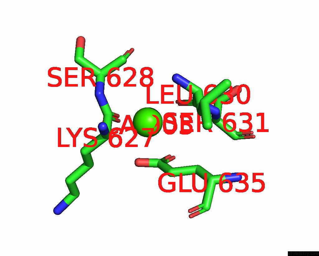
Mono view
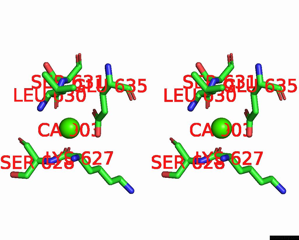
Stereo pair view
|
A full contact list of Calcium with other atoms in the Ca binding
site number 3 of The Pdz Domain of MAGI1_2 Complexed with the Pdz-Binding Motif of HPV35-E6 within 5.0Å range:
probe
|
atom
|
residue
|
distance (Å)
|
B
|
Occ
|
A:Ca903
b:254.8
occ:1.00
|
O
|
A:LYS627
|
2.5
|
115.7
|
1.0
|
O
|
A:LEU630
|
2.5
|
110.3
|
1.0
|
OE1
|
A:GLU635
|
2.6
|
136.7
|
1.0
|
OE2
|
A:GLU635
|
2.7
|
136.7
|
1.0
|
CD
|
A:GLU635
|
2.8
|
130.5
|
1.0
|
C
|
A:LEU630
|
3.7
|
106.8
|
1.0
|
C
|
A:LYS627
|
3.7
|
111.4
|
1.0
|
CG
|
A:GLU635
|
4.1
|
116.0
|
1.0
|
CA
|
A:SER631
|
4.2
|
108.2
|
1.0
|
N
|
A:SER631
|
4.4
|
106.9
|
1.0
|
CA
|
A:SER628
|
4.5
|
106.2
|
1.0
|
N
|
A:SER628
|
4.5
|
106.3
|
1.0
|
CA
|
A:LYS627
|
4.6
|
113.8
|
1.0
|
O
|
A:SER628
|
4.6
|
117.0
|
1.0
|
C
|
A:SER628
|
4.6
|
111.7
|
1.0
|
CB
|
A:SER631
|
4.6
|
111.9
|
1.0
|
CB
|
A:GLU635
|
4.7
|
104.4
|
1.0
|
N
|
A:LEU630
|
4.7
|
106.7
|
1.0
|
CG
|
A:LYS627
|
4.7
|
123.5
|
1.0
|
CA
|
A:LEU630
|
4.8
|
101.5
|
1.0
|
|
Calcium binding site 4 out
of 8 in 7p71
Go back to
Calcium Binding Sites List in 7p71
Calcium binding site 4 out
of 8 in the The Pdz Domain of MAGI1_2 Complexed with the Pdz-Binding Motif of HPV35-E6
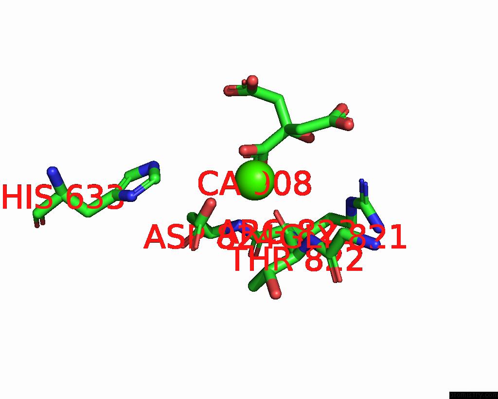
Mono view
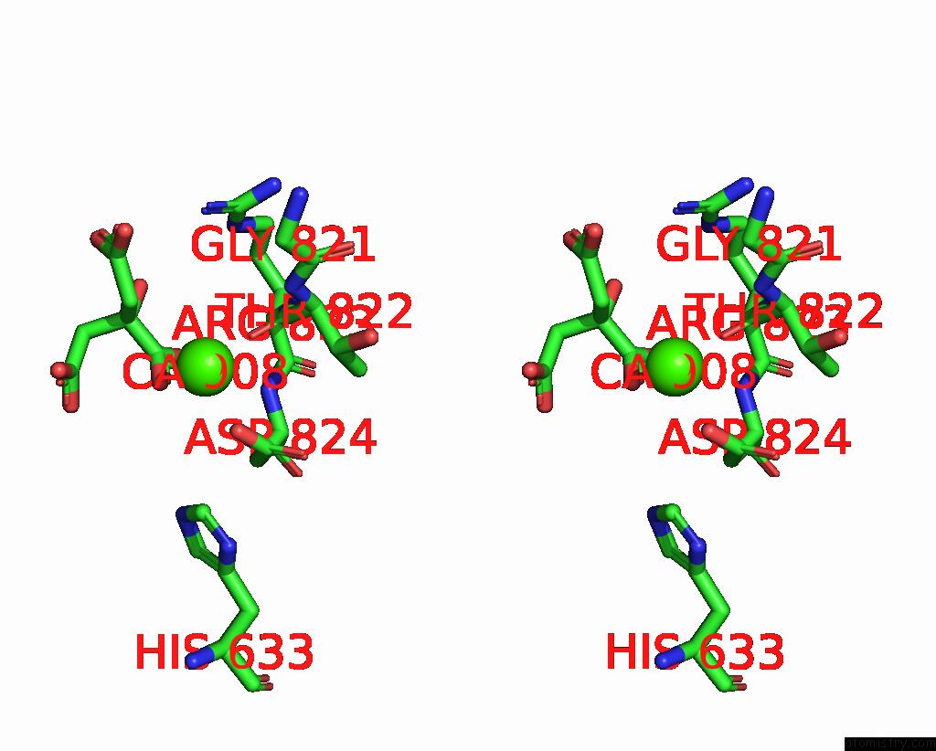
Stereo pair view
|
A full contact list of Calcium with other atoms in the Ca binding
site number 4 of The Pdz Domain of MAGI1_2 Complexed with the Pdz-Binding Motif of HPV35-E6 within 5.0Å range:
probe
|
atom
|
residue
|
distance (Å)
|
B
|
Occ
|
A:Ca908
b:164.3
occ:1.00
|
O6
|
A:CIT907
|
2.6
|
149.3
|
1.0
|
OD2
|
A:ASP824
|
2.6
|
85.2
|
1.0
|
O
|
A:THR822
|
2.7
|
85.9
|
1.0
|
C6
|
A:CIT907
|
3.4
|
149.9
|
1.0
|
C4
|
A:CIT907
|
3.4
|
153.3
|
1.0
|
N
|
A:THR822
|
3.7
|
76.3
|
1.0
|
C
|
A:THR822
|
3.8
|
71.5
|
1.0
|
CG
|
A:ASP824
|
3.8
|
78.9
|
1.0
|
C3
|
A:CIT907
|
4.1
|
151.1
|
1.0
|
CA
|
A:THR822
|
4.1
|
73.0
|
1.0
|
O5
|
A:CIT907
|
4.1
|
149.6
|
1.0
|
CB
|
A:THR822
|
4.2
|
72.8
|
1.0
|
OD1
|
A:ASP824
|
4.4
|
77.6
|
1.0
|
CE1
|
A:HIS633
|
4.5
|
93.2
|
1.0
|
C5
|
A:CIT907
|
4.7
|
155.2
|
1.0
|
C
|
A:GLY821
|
4.8
|
75.9
|
1.0
|
O2
|
A:CIT907
|
4.8
|
143.4
|
1.0
|
CB
|
A:ASP824
|
4.8
|
72.2
|
1.0
|
N
|
A:ASP824
|
4.8
|
66.1
|
1.0
|
NE2
|
A:HIS633
|
4.9
|
89.8
|
1.0
|
O4
|
A:CIT907
|
4.9
|
155.1
|
1.0
|
C1
|
A:CIT907
|
4.9
|
144.6
|
1.0
|
CA
|
A:GLY821
|
4.9
|
70.0
|
1.0
|
N
|
A:ARG823
|
5.0
|
64.9
|
1.0
|
|
Calcium binding site 5 out
of 8 in 7p71
Go back to
Calcium Binding Sites List in 7p71
Calcium binding site 5 out
of 8 in the The Pdz Domain of MAGI1_2 Complexed with the Pdz-Binding Motif of HPV35-E6
Mono view
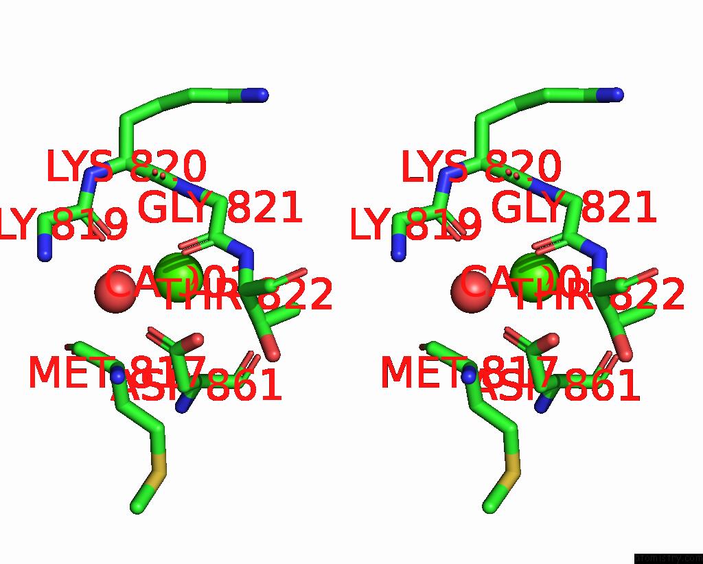
Stereo pair view
|
A full contact list of Calcium with other atoms in the Ca binding
site number 5 of The Pdz Domain of MAGI1_2 Complexed with the Pdz-Binding Motif of HPV35-E6 within 5.0Å range:
probe
|
atom
|
residue
|
distance (Å)
|
B
|
Occ
|
B:Ca901
b:180.3
occ:1.00
|
O
|
B:HOH1001
|
2.4
|
69.5
|
1.0
|
O
|
B:GLY821
|
2.4
|
110.3
|
1.0
|
OD2
|
B:ASP861
|
2.4
|
118.1
|
1.0
|
OD1
|
B:ASP861
|
2.4
|
123.7
|
1.0
|
CG
|
B:ASP861
|
2.7
|
115.2
|
1.0
|
C
|
B:GLY821
|
2.7
|
105.6
|
1.0
|
O
|
B:GLY819
|
3.1
|
84.8
|
1.0
|
N
|
B:GLY821
|
3.3
|
107.4
|
1.0
|
CA
|
B:GLY821
|
3.3
|
108.4
|
1.0
|
N
|
B:THR822
|
3.5
|
98.5
|
1.0
|
C
|
B:LYS820
|
3.9
|
106.5
|
1.0
|
CA
|
B:THR822
|
4.0
|
88.0
|
1.0
|
CB
|
B:THR822
|
4.0
|
90.8
|
1.0
|
CB
|
B:ASP861
|
4.2
|
103.5
|
1.0
|
C
|
B:GLY819
|
4.3
|
91.2
|
1.0
|
O
|
B:LYS820
|
4.5
|
111.6
|
1.0
|
CG2
|
B:THR822
|
4.5
|
105.5
|
1.0
|
CA
|
B:LYS820
|
4.6
|
101.5
|
1.0
|
O
|
B:ASP861
|
4.6
|
99.5
|
1.0
|
CA
|
B:ASP861
|
4.7
|
95.8
|
1.0
|
CA
|
B:MET817
|
4.8
|
71.5
|
1.0
|
O
|
B:MET817
|
4.8
|
76.8
|
1.0
|
C
|
B:MET817
|
5.0
|
77.1
|
1.0
|
N
|
B:LYS820
|
5.0
|
102.4
|
1.0
|
|
Calcium binding site 6 out
of 8 in 7p71
Go back to
Calcium Binding Sites List in 7p71
Calcium binding site 6 out
of 8 in the The Pdz Domain of MAGI1_2 Complexed with the Pdz-Binding Motif of HPV35-E6
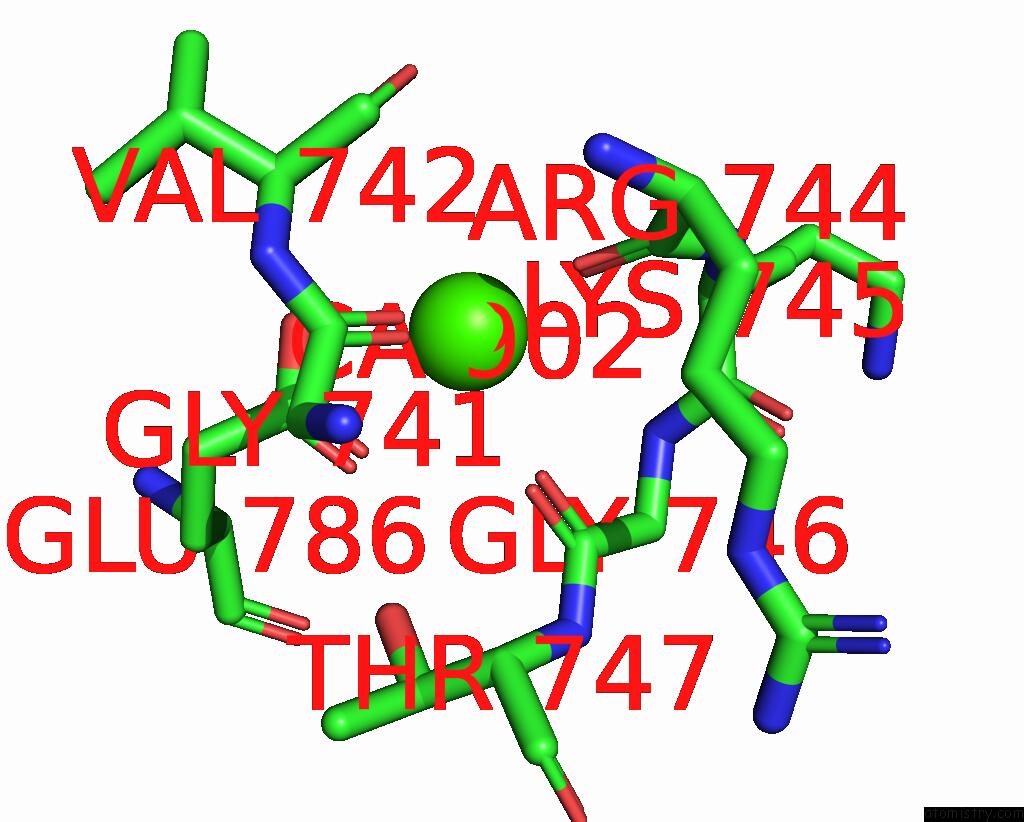
Mono view
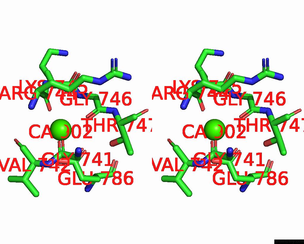
Stereo pair view
|
A full contact list of Calcium with other atoms in the Ca binding
site number 6 of The Pdz Domain of MAGI1_2 Complexed with the Pdz-Binding Motif of HPV35-E6 within 5.0Å range:
probe
|
atom
|
residue
|
distance (Å)
|
B
|
Occ
|
B:Ca902
b:151.5
occ:1.00
|
O
|
B:GLY741
|
2.4
|
107.7
|
1.0
|
O
|
B:ARG744
|
2.5
|
122.0
|
1.0
|
OE2
|
B:GLU786
|
2.6
|
144.0
|
1.0
|
OE1
|
B:GLU786
|
2.6
|
146.6
|
1.0
|
O
|
B:GLY746
|
2.7
|
87.6
|
1.0
|
CD
|
B:GLU786
|
3.0
|
137.6
|
1.0
|
N
|
B:GLY746
|
3.4
|
99.2
|
1.0
|
C
|
B:GLY741
|
3.6
|
88.2
|
1.0
|
C
|
B:ARG744
|
3.6
|
120.0
|
1.0
|
C
|
B:GLY746
|
3.7
|
93.0
|
1.0
|
CA
|
B:LYS745
|
3.8
|
107.7
|
1.0
|
C
|
B:LYS745
|
4.0
|
100.3
|
1.0
|
CA
|
B:GLY746
|
4.1
|
98.3
|
1.0
|
O
|
B:VAL742
|
4.2
|
107.8
|
1.0
|
CA
|
B:VAL742
|
4.2
|
94.1
|
1.0
|
N
|
B:LYS745
|
4.2
|
115.6
|
1.0
|
C
|
B:VAL742
|
4.3
|
102.1
|
1.0
|
N
|
B:VAL742
|
4.3
|
93.1
|
1.0
|
CG
|
B:GLU786
|
4.5
|
121.1
|
1.0
|
OG1
|
B:THR747
|
4.5
|
99.3
|
1.0
|
CA
|
B:GLY741
|
4.6
|
76.2
|
1.0
|
N
|
B:THR747
|
4.9
|
97.2
|
1.0
|
CA
|
B:ARG744
|
4.9
|
121.0
|
1.0
|
N
|
B:ARG744
|
4.9
|
109.6
|
1.0
|
O
|
B:LYS745
|
5.0
|
98.3
|
1.0
|
|
Calcium binding site 7 out
of 8 in 7p71
Go back to
Calcium Binding Sites List in 7p71
Calcium binding site 7 out
of 8 in the The Pdz Domain of MAGI1_2 Complexed with the Pdz-Binding Motif of HPV35-E6
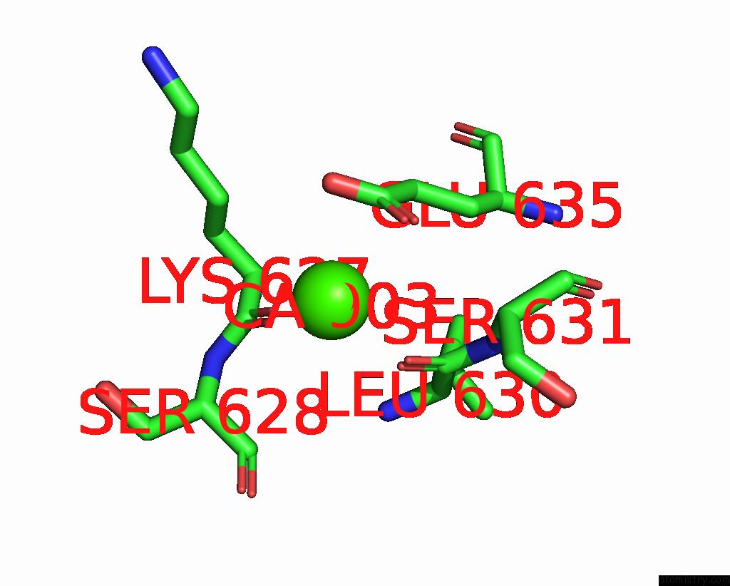
Mono view
Stereo pair view
|
A full contact list of Calcium with other atoms in the Ca binding
site number 7 of The Pdz Domain of MAGI1_2 Complexed with the Pdz-Binding Motif of HPV35-E6 within 5.0Å range:
probe
|
atom
|
residue
|
distance (Å)
|
B
|
Occ
|
B:Ca903
b:152.2
occ:1.00
|
O
|
B:LEU630
|
2.7
|
83.9
|
1.0
|
OE1
|
B:GLU635
|
2.7
|
96.4
|
1.0
|
O
|
B:LYS627
|
2.8
|
119.9
|
1.0
|
CD
|
B:GLU635
|
3.3
|
95.5
|
1.0
|
OE2
|
B:GLU635
|
3.3
|
98.1
|
1.0
|
C
|
B:LEU630
|
3.8
|
88.2
|
1.0
|
C
|
B:LYS627
|
4.0
|
114.4
|
1.0
|
CA
|
B:SER631
|
4.1
|
98.0
|
1.0
|
CB
|
B:SER631
|
4.4
|
106.0
|
1.0
|
N
|
B:SER631
|
4.4
|
94.6
|
1.0
|
CG
|
B:GLU635
|
4.6
|
91.0
|
1.0
|
CG
|
B:LYS627
|
4.7
|
113.8
|
1.0
|
CA
|
B:LYS627
|
4.9
|
111.1
|
1.0
|
N
|
B:LEU630
|
4.9
|
95.9
|
1.0
|
CA
|
B:SER628
|
4.9
|
104.9
|
1.0
|
N
|
B:SER628
|
4.9
|
108.7
|
1.0
|
CA
|
B:LEU630
|
5.0
|
91.6
|
1.0
|
|
Calcium binding site 8 out
of 8 in 7p71
Go back to
Calcium Binding Sites List in 7p71
Calcium binding site 8 out
of 8 in the The Pdz Domain of MAGI1_2 Complexed with the Pdz-Binding Motif of HPV35-E6
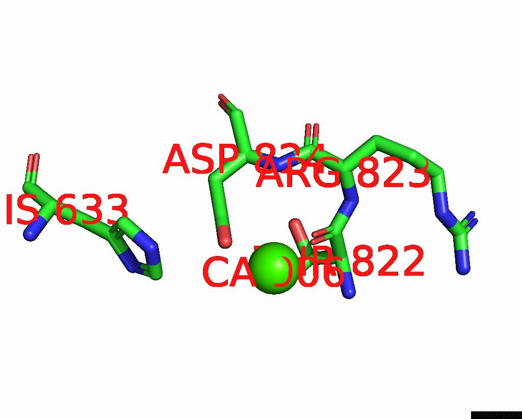
Mono view
Stereo pair view
|
A full contact list of Calcium with other atoms in the Ca binding
site number 8 of The Pdz Domain of MAGI1_2 Complexed with the Pdz-Binding Motif of HPV35-E6 within 5.0Å range:
probe
|
atom
|
residue
|
distance (Å)
|
B
|
Occ
|
B:Ca906
b:122.0
occ:1.00
|
O
|
B:THR822
|
2.6
|
83.9
|
1.0
|
OD2
|
B:ASP824
|
3.0
|
89.4
|
1.0
|
C
|
B:THR822
|
3.8
|
77.9
|
1.0
|
CG
|
B:ASP824
|
4.0
|
84.1
|
1.0
|
N
|
B:ASP824
|
4.4
|
82.8
|
1.0
|
CG2
|
B:THR822
|
4.4
|
105.5
|
1.0
|
N
|
B:THR822
|
4.5
|
98.5
|
1.0
|
NE2
|
B:HIS633
|
4.6
|
82.5
|
1.0
|
CB
|
B:ASP824
|
4.7
|
82.2
|
1.0
|
CA
|
B:THR822
|
4.7
|
88.0
|
1.0
|
CE1
|
B:HIS633
|
4.7
|
84.3
|
1.0
|
N
|
B:ARG823
|
4.8
|
73.9
|
1.0
|
CA
|
B:ARG823
|
4.9
|
79.6
|
1.0
|
OD1
|
B:ASP824
|
4.9
|
85.8
|
1.0
|
|
Reference:
G.Gogl,
A.Cousido-Siah,
G.Trave.
The Pdz Domain of MAGI1_2 Complexed with the Pdz-Binding Motif of HPV35-E6 To Be Published.
Page generated: Fri Jul 19 02:44:47 2024
|