Calcium »
PDB 7or8-7p9r »
7p72 »
Calcium in PDB 7p72: The Pdz Domain of SNX27 Complexed with the Pdz-Binding Motif of Mers-E
Protein crystallography data
The structure of The Pdz Domain of SNX27 Complexed with the Pdz-Binding Motif of Mers-E, PDB code: 7p72
was solved by
G.Gogl,
A.Cousido-Siah,
G.Trave,
with X-Ray Crystallography technique. A brief refinement statistics is given in the table below:
| Resolution Low / High (Å) | 44.99 / 2.15 |
| Space group | P 21 21 21 |
| Cell size a, b, c (Å), α, β, γ (°) | 61.17, 83.95, 106.59, 90, 90, 90 |
| R / Rfree (%) | 19.9 / 24 |
Calcium Binding Sites:
The binding sites of Calcium atom in the The Pdz Domain of SNX27 Complexed with the Pdz-Binding Motif of Mers-E
(pdb code 7p72). This binding sites where shown within
5.0 Angstroms radius around Calcium atom.
In total 5 binding sites of Calcium where determined in the The Pdz Domain of SNX27 Complexed with the Pdz-Binding Motif of Mers-E, PDB code: 7p72:
Jump to Calcium binding site number: 1; 2; 3; 4; 5;
In total 5 binding sites of Calcium where determined in the The Pdz Domain of SNX27 Complexed with the Pdz-Binding Motif of Mers-E, PDB code: 7p72:
Jump to Calcium binding site number: 1; 2; 3; 4; 5;
Calcium binding site 1 out of 5 in 7p72
Go back to
Calcium binding site 1 out
of 5 in the The Pdz Domain of SNX27 Complexed with the Pdz-Binding Motif of Mers-E
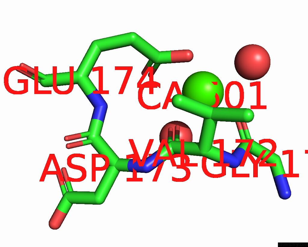
Mono view

Stereo pair view
Mono view
Stereo pair view
A full contact list of Calcium with other atoms in the Ca binding
site number 1 of The Pdz Domain of SNX27 Complexed with the Pdz-Binding Motif of Mers-E within 5.0Å range:
|
Calcium binding site 2 out of 5 in 7p72
Go back to
Calcium binding site 2 out
of 5 in the The Pdz Domain of SNX27 Complexed with the Pdz-Binding Motif of Mers-E

Mono view
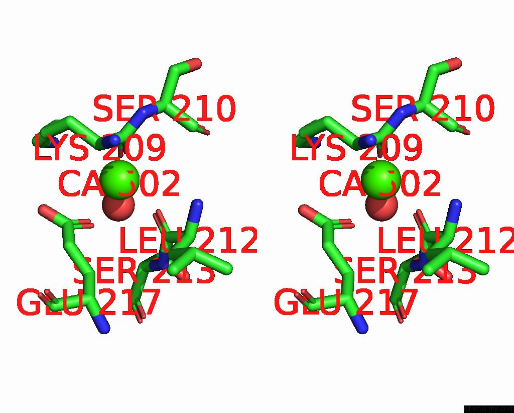
Stereo pair view
Mono view
Stereo pair view
A full contact list of Calcium with other atoms in the Ca binding
site number 2 of The Pdz Domain of SNX27 Complexed with the Pdz-Binding Motif of Mers-E within 5.0Å range:
|
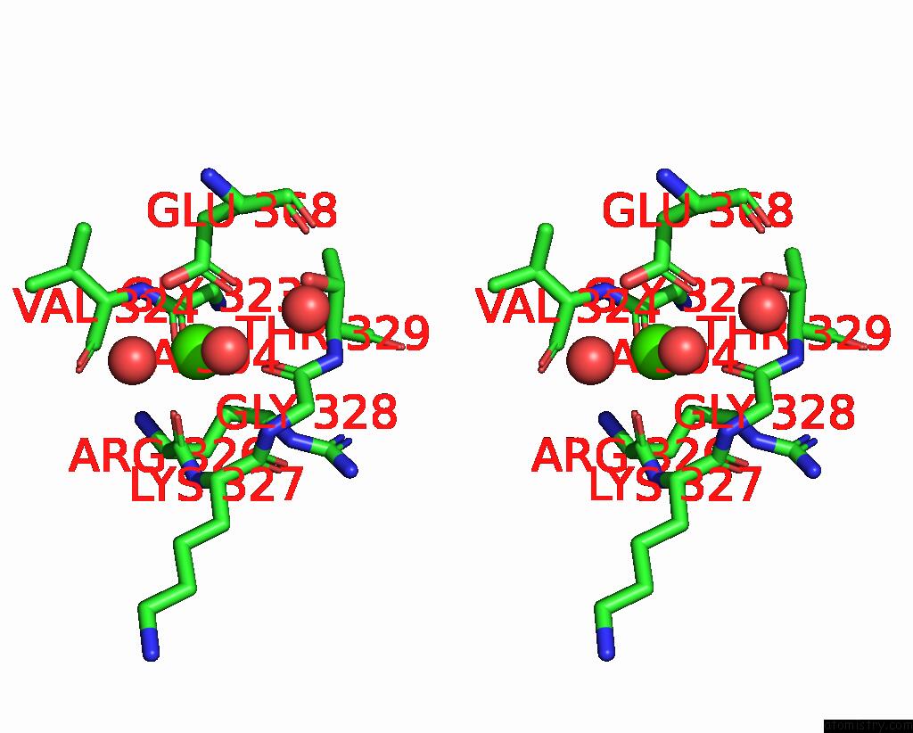
Calcium binding site 3 out of 5 in 7p72
Go back to
Calcium binding site 3 out
of 5 in the The Pdz Domain of SNX27 Complexed with the Pdz-Binding Motif of Mers-E

Mono view

Stereo pair view
Mono view
Stereo pair view
A full contact list of Calcium with other atoms in the Ca binding
site number 3 of The Pdz Domain of SNX27 Complexed with the Pdz-Binding Motif of Mers-E within 5.0Å range:
|
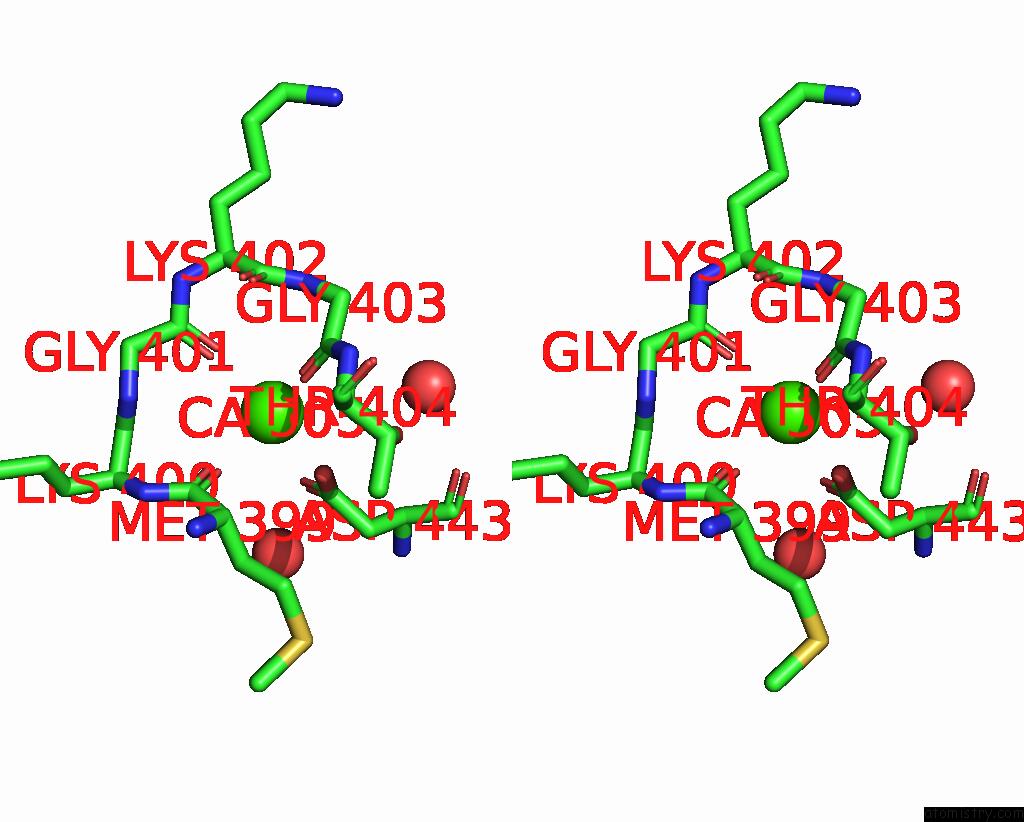
Calcium binding site 4 out of 5 in 7p72
Go back to
Calcium binding site 4 out
of 5 in the The Pdz Domain of SNX27 Complexed with the Pdz-Binding Motif of Mers-E

Mono view
Stereo pair view
Mono view
Stereo pair view
A full contact list of Calcium with other atoms in the Ca binding
site number 4 of The Pdz Domain of SNX27 Complexed with the Pdz-Binding Motif of Mers-E within 5.0Å range:
|
Calcium binding site 5 out of 5 in 7p72
Go back to
Calcium binding site 5 out
of 5 in the The Pdz Domain of SNX27 Complexed with the Pdz-Binding Motif of Mers-E

Mono view
Stereo pair view
Mono view
Stereo pair view
A full contact list of Calcium with other atoms in the Ca binding
site number 5 of The Pdz Domain of SNX27 Complexed with the Pdz-Binding Motif of Mers-E within 5.0Å range:
|
Reference:
G.Gogl,
A.Cousido-Siah,
G.Trave.
The Pdz Domain of SNX27 Complexed with the Pdz-Binding Motif of Mers-E To Be Published.
Page generated: Thu Jul 10 00:01:44 2025
Last articles
Cl in 8EYACl in 8EZF
Cl in 8EWO
Cl in 8EY0
Cl in 8EWA
Cl in 8EW1
Cl in 8EVO
Cl in 8ESX
Cl in 8ESY
Cl in 8EQM