Calcium »
PDB 7or8-7p9r »
7p74 »
Calcium in PDB 7p74: The Pdz Domain of SYNJ2BP Complexed with the Phosphorylated Pdz- Binding Motif of RSK1
Enzymatic activity of The Pdz Domain of SYNJ2BP Complexed with the Phosphorylated Pdz- Binding Motif of RSK1
All present enzymatic activity of The Pdz Domain of SYNJ2BP Complexed with the Phosphorylated Pdz- Binding Motif of RSK1:
2.7.11.1;
2.7.11.1;
Protein crystallography data
The structure of The Pdz Domain of SYNJ2BP Complexed with the Phosphorylated Pdz- Binding Motif of RSK1, PDB code: 7p74
was solved by
G.Gogl,
A.Cousido-Siah,
G.Trave,
with X-Ray Crystallography technique. A brief refinement statistics is given in the table below:
| Resolution Low / High (Å) | 46.71 / 1.90 |
| Space group | P 21 21 21 |
| Cell size a, b, c (Å), α, β, γ (°) | 59.62, 75.15, 108.46, 90, 90, 90 |
| R / Rfree (%) | 18.1 / 21.1 |
Calcium Binding Sites:
The binding sites of Calcium atom in the The Pdz Domain of SYNJ2BP Complexed with the Phosphorylated Pdz- Binding Motif of RSK1
(pdb code 7p74). This binding sites where shown within
5.0 Angstroms radius around Calcium atom.
In total 4 binding sites of Calcium where determined in the The Pdz Domain of SYNJ2BP Complexed with the Phosphorylated Pdz- Binding Motif of RSK1, PDB code: 7p74:
Jump to Calcium binding site number: 1; 2; 3; 4;
In total 4 binding sites of Calcium where determined in the The Pdz Domain of SYNJ2BP Complexed with the Phosphorylated Pdz- Binding Motif of RSK1, PDB code: 7p74:
Jump to Calcium binding site number: 1; 2; 3; 4;
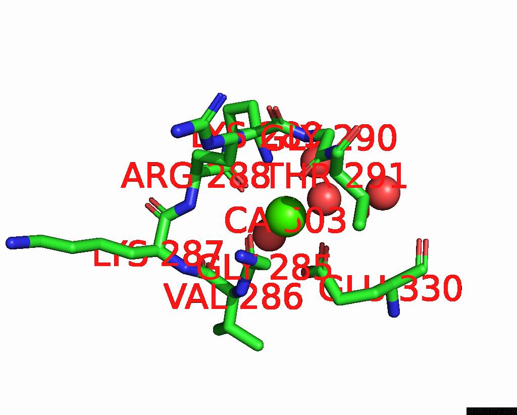
Calcium binding site 1 out of 4 in 7p74
Go back to
Calcium binding site 1 out
of 4 in the The Pdz Domain of SYNJ2BP Complexed with the Phosphorylated Pdz- Binding Motif of RSK1
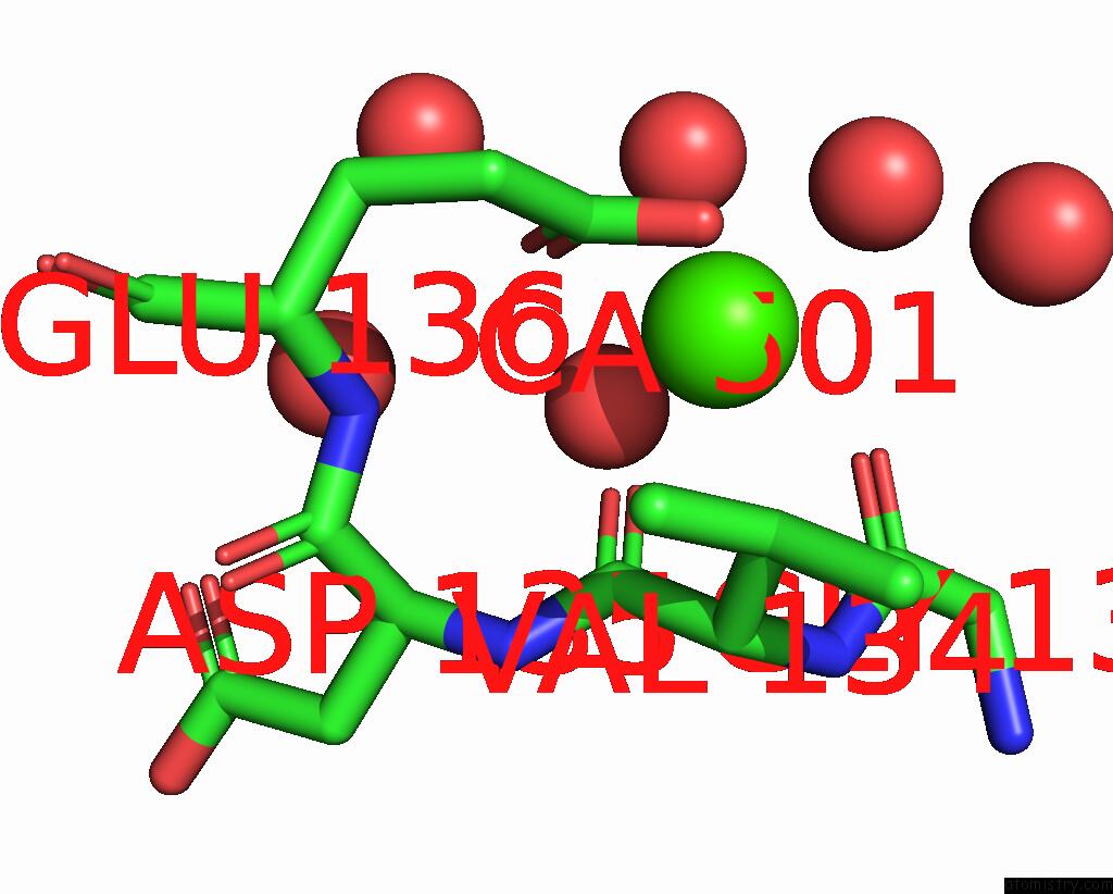
Mono view
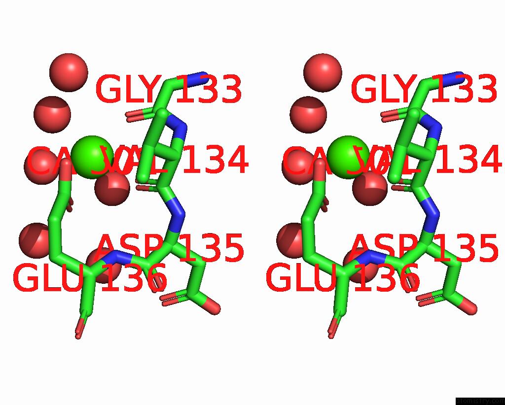
Stereo pair view
Mono view
Stereo pair view
A full contact list of Calcium with other atoms in the Ca binding
site number 1 of The Pdz Domain of SYNJ2BP Complexed with the Phosphorylated Pdz- Binding Motif of RSK1 within 5.0Å range:
|
Calcium binding site 2 out of 4 in 7p74
Go back to
Calcium binding site 2 out
of 4 in the The Pdz Domain of SYNJ2BP Complexed with the Phosphorylated Pdz- Binding Motif of RSK1
Mono view
Stereo pair view
Mono view
Stereo pair view
A full contact list of Calcium with other atoms in the Ca binding
site number 2 of The Pdz Domain of SYNJ2BP Complexed with the Phosphorylated Pdz- Binding Motif of RSK1 within 5.0Å range:
|
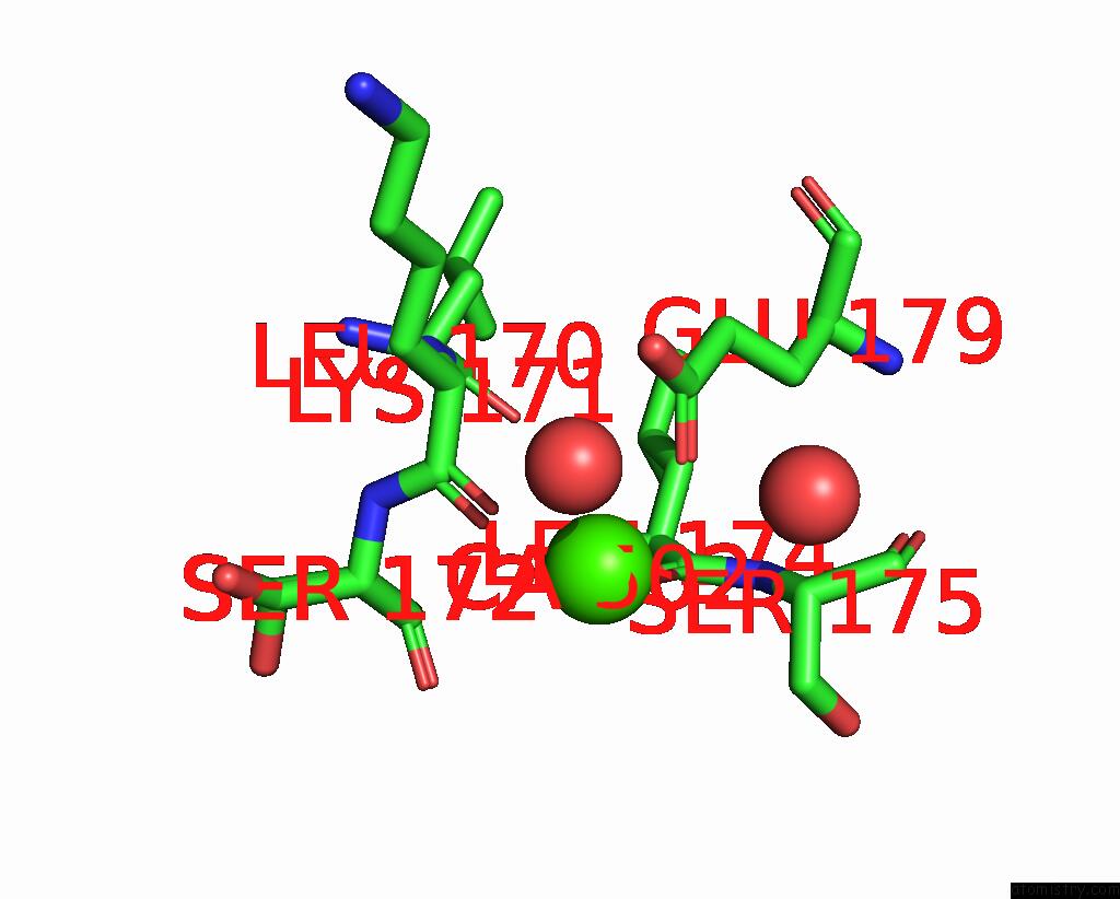
Calcium binding site 3 out of 4 in 7p74
Go back to
Calcium binding site 3 out
of 4 in the The Pdz Domain of SYNJ2BP Complexed with the Phosphorylated Pdz- Binding Motif of RSK1
Mono view

Stereo pair view
Mono view
Stereo pair view
A full contact list of Calcium with other atoms in the Ca binding
site number 3 of The Pdz Domain of SYNJ2BP Complexed with the Phosphorylated Pdz- Binding Motif of RSK1 within 5.0Å range:
|
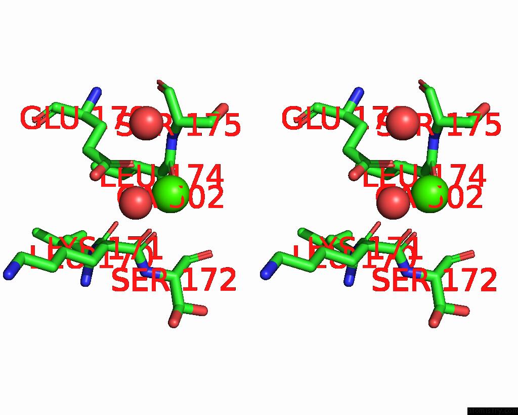
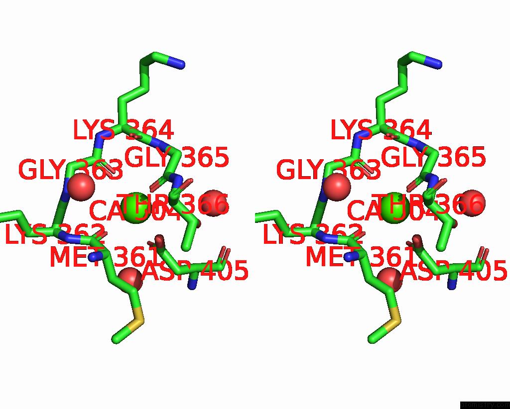
Calcium binding site 4 out of 4 in 7p74
Go back to
Calcium binding site 4 out
of 4 in the The Pdz Domain of SYNJ2BP Complexed with the Phosphorylated Pdz- Binding Motif of RSK1

Mono view
Stereo pair view
Mono view
Stereo pair view
A full contact list of Calcium with other atoms in the Ca binding
site number 4 of The Pdz Domain of SYNJ2BP Complexed with the Phosphorylated Pdz- Binding Motif of RSK1 within 5.0Å range:
|
Reference:
G.Gogl,
A.Cousido-Siah,
G.Trave.
The Pdz Domain of SYNJ2BP Complexed with the Phosphorylated Pdz-Binding Motif of RSK1 To Be Published.
Page generated: Thu Jul 10 00:02:50 2025
Last articles
F in 7QIRF in 7QI8
F in 7QC5
F in 7QG3
F in 7QHN
F in 7Q6P
F in 7QBZ
F in 7QC0
F in 7QAV
F in 7QA0