Calcium in PDB 7pc5: The Third Pdz Domain of PDZD7 Complexed with the Pdz-Binding Motif of EXOC4
Protein crystallography data
The structure of The Third Pdz Domain of PDZD7 Complexed with the Pdz-Binding Motif of EXOC4, PDB code: 7pc5
was solved by
A.Cousido-Siah,
G.Trave,
G.Gogl,
with X-Ray Crystallography technique. A brief refinement statistics is given in the table below:
Resolution Low / High (Å)
|
42.75 /
1.70
|
Space group
|
P 2 21 21
|
Cell size a, b, c (Å), α, β, γ (°)
|
54.11,
60.35,
181.68,
90,
90,
90
|
R / Rfree (%)
|
16.6 /
18.8
|
Calcium Binding Sites:
The binding sites of Calcium atom in the The Third Pdz Domain of PDZD7 Complexed with the Pdz-Binding Motif of EXOC4
(pdb code 7pc5). This binding sites where shown within
5.0 Angstroms radius around Calcium atom.
In total 5 binding sites of Calcium where determined in the
The Third Pdz Domain of PDZD7 Complexed with the Pdz-Binding Motif of EXOC4, PDB code: 7pc5:
Jump to Calcium binding site number:
1;
2;
3;
4;
5;
Calcium binding site 1 out
of 5 in 7pc5
Go back to
Calcium Binding Sites List in 7pc5
Calcium binding site 1 out
of 5 in the The Third Pdz Domain of PDZD7 Complexed with the Pdz-Binding Motif of EXOC4
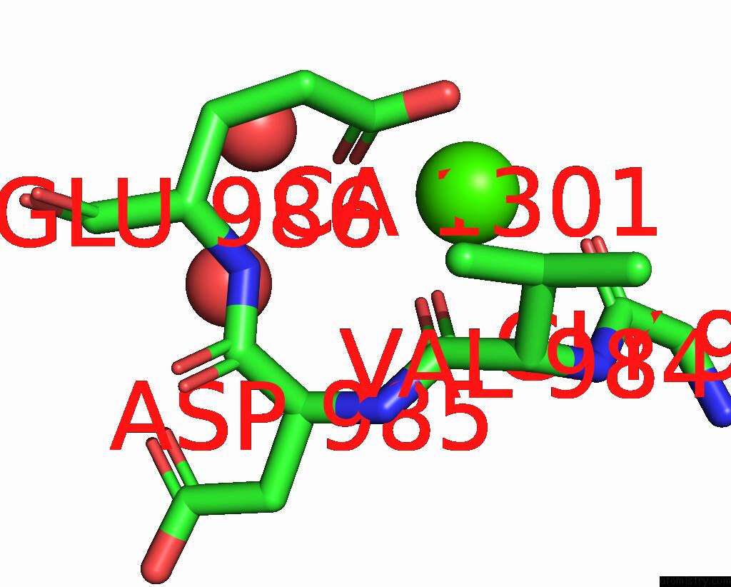
Mono view
Stereo pair view
|
A full contact list of Calcium with other atoms in the Ca binding
site number 1 of The Third Pdz Domain of PDZD7 Complexed with the Pdz-Binding Motif of EXOC4 within 5.0Å range:
probe
|
atom
|
residue
|
distance (Å)
|
B
|
Occ
|
A:Ca1301
b:58.6
occ:1.00
|
OE1
|
A:GLU986
|
2.4
|
47.6
|
1.0
|
O
|
A:VAL984
|
2.5
|
36.9
|
1.0
|
O
|
A:GLY983
|
2.6
|
39.4
|
1.0
|
OE2
|
A:GLU986
|
2.7
|
38.9
|
1.0
|
CD
|
A:GLU986
|
2.9
|
41.8
|
1.0
|
C
|
A:GLY983
|
3.5
|
39.9
|
1.0
|
C
|
A:VAL984
|
3.7
|
29.5
|
1.0
|
N
|
A:VAL984
|
4.2
|
33.3
|
1.0
|
O
|
A:HOH1497
|
4.3
|
44.0
|
1.0
|
CA
|
A:VAL984
|
4.3
|
29.3
|
1.0
|
CB
|
A:VAL984
|
4.4
|
29.5
|
1.0
|
CG
|
A:GLU986
|
4.4
|
28.4
|
1.0
|
CA
|
A:GLY983
|
4.4
|
41.9
|
1.0
|
O
|
A:HOH1728
|
4.6
|
45.4
|
1.0
|
N
|
A:GLU986
|
4.7
|
25.9
|
1.0
|
N
|
A:ASP985
|
4.8
|
27.7
|
1.0
|
|
Calcium binding site 2 out
of 5 in 7pc5
Go back to
Calcium Binding Sites List in 7pc5
Calcium binding site 2 out
of 5 in the The Third Pdz Domain of PDZD7 Complexed with the Pdz-Binding Motif of EXOC4
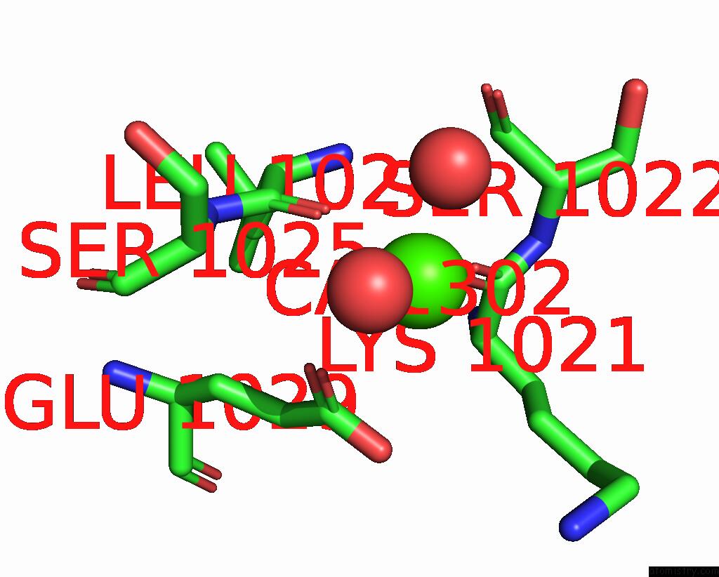
Mono view
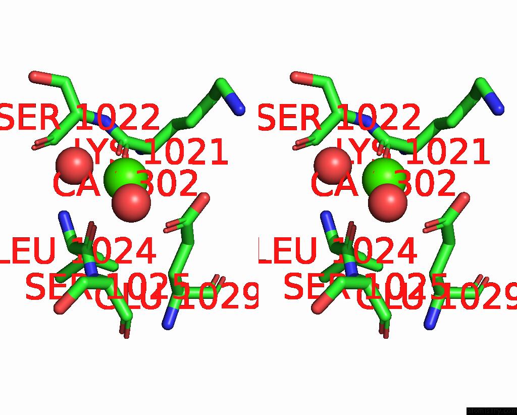
Stereo pair view
|
A full contact list of Calcium with other atoms in the Ca binding
site number 2 of The Third Pdz Domain of PDZD7 Complexed with the Pdz-Binding Motif of EXOC4 within 5.0Å range:
probe
|
atom
|
residue
|
distance (Å)
|
B
|
Occ
|
A:Ca1302
b:64.1
occ:1.00
|
O
|
A:HOH1733
|
2.2
|
58.7
|
1.0
|
O
|
A:HOH1698
|
2.2
|
50.3
|
1.0
|
O
|
A:LYS1021
|
2.5
|
35.5
|
1.0
|
O
|
A:LEU1024
|
2.7
|
29.5
|
1.0
|
OE1
|
A:GLU1029
|
2.7
|
39.8
|
1.0
|
CD
|
A:GLU1029
|
3.4
|
41.6
|
1.0
|
OE2
|
A:GLU1029
|
3.6
|
45.6
|
1.0
|
C
|
A:LYS1021
|
3.7
|
35.6
|
1.0
|
C
|
A:LEU1024
|
3.8
|
29.1
|
1.0
|
CA
|
A:SER1025
|
4.3
|
28.7
|
1.0
|
CG
|
A:LYS1021
|
4.5
|
41.2
|
1.0
|
CA
|
A:SER1022
|
4.5
|
30.3
|
1.0
|
O
|
A:SER1022
|
4.5
|
37.1
|
1.0
|
CA
|
A:LYS1021
|
4.6
|
33.0
|
1.0
|
N
|
A:SER1025
|
4.6
|
26.7
|
1.0
|
N
|
A:SER1022
|
4.6
|
29.0
|
1.0
|
CB
|
A:SER1025
|
4.6
|
35.2
|
1.0
|
CG
|
A:GLU1029
|
4.6
|
27.8
|
1.0
|
C
|
A:SER1022
|
4.6
|
37.2
|
1.0
|
N
|
A:LEU1024
|
4.8
|
25.9
|
1.0
|
CA
|
A:LEU1024
|
4.9
|
25.8
|
1.0
|
|
Calcium binding site 3 out
of 5 in 7pc5
Go back to
Calcium Binding Sites List in 7pc5
Calcium binding site 3 out
of 5 in the The Third Pdz Domain of PDZD7 Complexed with the Pdz-Binding Motif of EXOC4
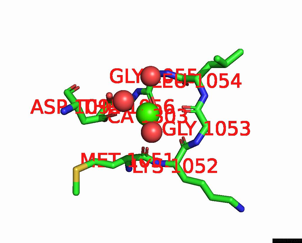
Mono view
Stereo pair view
|
A full contact list of Calcium with other atoms in the Ca binding
site number 3 of The Third Pdz Domain of PDZD7 Complexed with the Pdz-Binding Motif of EXOC4 within 5.0Å range:
probe
|
atom
|
residue
|
distance (Å)
|
B
|
Occ
|
A:Ca1303
b:50.9
occ:1.00
|
O
|
A:MET1051
|
2.6
|
37.1
|
1.0
|
O
|
A:GLY1055
|
2.7
|
34.0
|
1.0
|
OD1
|
A:ASP1095
|
2.7
|
64.1
|
1.0
|
O
|
A:GLY1053
|
2.7
|
34.5
|
1.0
|
O
|
A:HOH1442
|
3.0
|
42.2
|
1.0
|
O
|
A:HOH1751
|
3.3
|
61.5
|
1.0
|
C
|
A:MET1051
|
3.5
|
32.7
|
1.0
|
CG
|
A:ASP1095
|
3.5
|
48.5
|
1.0
|
OD2
|
A:ASP1095
|
3.5
|
51.3
|
1.0
|
C
|
A:GLY1055
|
3.5
|
46.8
|
1.0
|
C
|
A:GLY1053
|
3.8
|
35.9
|
1.0
|
N
|
A:GLY1055
|
3.8
|
41.9
|
1.0
|
O
|
A:LYS1052
|
4.1
|
37.6
|
1.0
|
C
|
A:LYS1052
|
4.1
|
37.7
|
1.0
|
CA
|
A:GLY1055
|
4.1
|
41.4
|
1.0
|
O
|
A:HOH1597
|
4.1
|
44.2
|
1.0
|
CA
|
A:MET1051
|
4.2
|
24.9
|
1.0
|
N
|
A:LYS1052
|
4.2
|
32.0
|
1.0
|
N
|
A:GLY1053
|
4.3
|
34.4
|
1.0
|
OG1
|
A:THR1056
|
4.3
|
44.2
|
1.0
|
C
|
A:LEU1054
|
4.3
|
44.6
|
1.0
|
CA
|
A:LYS1052
|
4.5
|
35.8
|
1.0
|
N
|
A:THR1056
|
4.5
|
38.2
|
1.0
|
CA
|
A:GLY1053
|
4.7
|
32.4
|
1.0
|
N
|
A:LEU1054
|
4.7
|
29.3
|
1.0
|
CA
|
A:LEU1054
|
4.7
|
28.5
|
1.0
|
CA
|
A:THR1056
|
4.8
|
31.7
|
1.0
|
CB
|
A:MET1051
|
4.9
|
29.3
|
1.0
|
O
|
A:LEU1054
|
4.9
|
44.0
|
1.0
|
CB
|
A:ASP1095
|
4.9
|
33.1
|
1.0
|
|
Calcium binding site 4 out
of 5 in 7pc5
Go back to
Calcium Binding Sites List in 7pc5
Calcium binding site 4 out
of 5 in the The Third Pdz Domain of PDZD7 Complexed with the Pdz-Binding Motif of EXOC4
Mono view
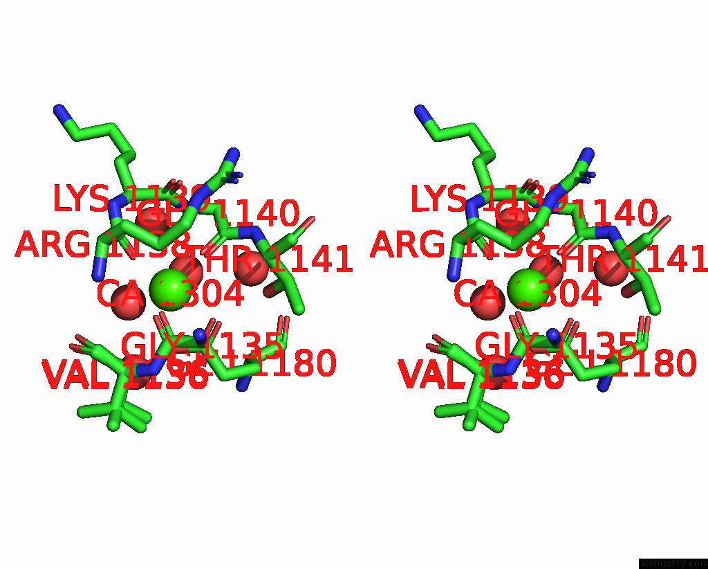
Stereo pair view
|
A full contact list of Calcium with other atoms in the Ca binding
site number 4 of The Third Pdz Domain of PDZD7 Complexed with the Pdz-Binding Motif of EXOC4 within 5.0Å range:
probe
|
atom
|
residue
|
distance (Å)
|
B
|
Occ
|
A:Ca1304
b:32.7
occ:1.00
|
O
|
A:GLY1140
|
2.3
|
29.4
|
1.0
|
O
|
A:ARG1138
|
2.3
|
29.4
|
1.0
|
O
|
A:GLY1135
|
2.3
|
29.4
|
1.0
|
OE1
|
A:GLU1180
|
2.4
|
26.4
|
1.0
|
O
|
A:HOH1662
|
2.5
|
42.7
|
1.0
|
O
|
A:HOH1747
|
2.5
|
38.0
|
1.0
|
OE2
|
A:GLU1180
|
2.6
|
30.9
|
1.0
|
CD
|
A:GLU1180
|
2.8
|
31.2
|
1.0
|
C
|
A:GLY1135
|
3.3
|
30.9
|
1.0
|
C
|
A:GLY1140
|
3.4
|
34.7
|
1.0
|
C
|
A:ARG1138
|
3.5
|
34.7
|
1.0
|
N
|
A:GLY1140
|
3.7
|
39.5
|
1.0
|
CA
|
A:GLY1140
|
4.0
|
38.0
|
1.0
|
CA
|
A:GLY1135
|
4.1
|
26.2
|
1.0
|
C
|
A:LYS1139
|
4.1
|
36.6
|
1.0
|
CA
|
A:VAL1136
|
4.1
|
27.0
|
0.3
|
N
|
A:VAL1136
|
4.1
|
27.2
|
0.3
|
N
|
A:VAL1136
|
4.2
|
26.0
|
0.7
|
OG1
|
A:THR1141
|
4.2
|
31.5
|
1.0
|
CA
|
A:VAL1136
|
4.2
|
25.9
|
0.7
|
N
|
A:ARG1138
|
4.3
|
37.8
|
1.0
|
CG
|
A:ARG1138
|
4.4
|
26.9
|
1.0
|
CG
|
A:GLU1180
|
4.4
|
27.2
|
1.0
|
O
|
A:HOH1523
|
4.4
|
36.0
|
1.0
|
CA
|
A:LYS1139
|
4.4
|
35.6
|
1.0
|
N
|
A:LYS1139
|
4.4
|
34.2
|
1.0
|
C
|
A:VAL1136
|
4.4
|
32.0
|
0.7
|
CA
|
A:ARG1138
|
4.4
|
37.4
|
1.0
|
N
|
A:THR1141
|
4.4
|
29.6
|
1.0
|
O
|
A:VAL1136
|
4.5
|
31.2
|
0.7
|
C
|
A:VAL1136
|
4.5
|
30.6
|
0.3
|
O
|
A:HOH1744
|
4.6
|
47.3
|
1.0
|
O
|
A:LYS1139
|
4.7
|
38.4
|
1.0
|
CA
|
A:THR1141
|
4.8
|
26.4
|
1.0
|
O
|
A:HOH1583
|
4.8
|
44.0
|
1.0
|
O
|
A:VAL1136
|
4.9
|
30.9
|
0.3
|
|
Calcium binding site 5 out
of 5 in 7pc5
Go back to
Calcium Binding Sites List in 7pc5
Calcium binding site 5 out
of 5 in the The Third Pdz Domain of PDZD7 Complexed with the Pdz-Binding Motif of EXOC4
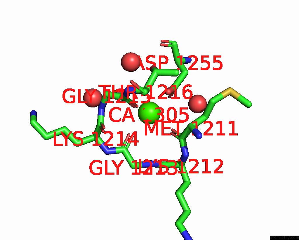
Mono view
Stereo pair view
|
A full contact list of Calcium with other atoms in the Ca binding
site number 5 of The Third Pdz Domain of PDZD7 Complexed with the Pdz-Binding Motif of EXOC4 within 5.0Å range:
probe
|
atom
|
residue
|
distance (Å)
|
B
|
Occ
|
A:Ca1305
b:20.1
occ:1.00
|
O
|
A:GLY1213
|
2.4
|
21.8
|
1.0
|
O
|
A:GLY1215
|
2.4
|
23.8
|
1.0
|
O
|
A:MET1211
|
2.4
|
20.1
|
1.0
|
OD1
|
A:ASP1255
|
2.5
|
20.5
|
1.0
|
OD2
|
A:ASP1255
|
2.5
|
21.4
|
1.0
|
CG
|
A:ASP1255
|
2.8
|
23.4
|
1.0
|
C
|
A:MET1211
|
3.4
|
19.0
|
1.0
|
C
|
A:GLY1215
|
3.5
|
26.2
|
1.0
|
C
|
A:GLY1213
|
3.6
|
24.3
|
1.0
|
N
|
A:GLY1215
|
4.0
|
25.2
|
1.0
|
CA
|
A:MET1211
|
4.0
|
16.7
|
1.0
|
N
|
A:GLY1213
|
4.1
|
23.3
|
1.0
|
CA
|
A:GLY1215
|
4.2
|
29.1
|
1.0
|
C
|
A:LYS1214
|
4.2
|
25.6
|
1.0
|
C
|
A:LYS1212
|
4.3
|
20.8
|
1.0
|
CB
|
A:ASP1255
|
4.3
|
21.4
|
1.0
|
O
|
A:HOH1647
|
4.3
|
30.6
|
1.0
|
OG1
|
A:THR1216
|
4.4
|
24.8
|
1.0
|
CA
|
A:GLY1213
|
4.4
|
23.6
|
1.0
|
N
|
A:LYS1212
|
4.5
|
20.4
|
1.0
|
N
|
A:THR1216
|
4.5
|
21.1
|
1.0
|
N
|
A:LYS1214
|
4.6
|
23.6
|
1.0
|
CB
|
A:MET1211
|
4.6
|
19.4
|
1.0
|
O
|
A:LYS1214
|
4.7
|
26.1
|
1.0
|
O
|
A:LYS1212
|
4.7
|
23.6
|
1.0
|
CA
|
A:LYS1214
|
4.7
|
23.8
|
1.0
|
CA
|
A:THR1216
|
4.8
|
22.6
|
1.0
|
O
|
A:HOH1556
|
4.8
|
20.9
|
1.0
|
CA
|
A:LYS1212
|
4.9
|
19.7
|
1.0
|
CA
|
A:ASP1255
|
5.0
|
21.4
|
1.0
|
O
|
A:HOH1614
|
5.0
|
33.4
|
1.0
|
|
Reference:
A.Cousido-Siah,
L.Carneiro,
C.Kostmann,
P.Ecsedi,
L.Nyitray,
G.Trave,
G.Gogl.
A Scalable Strategy to Solve Structures of Pdz Domains and Their Complexes. Acta Crystallogr D Struct V. 78 509 2022BIOL.
ISSN: ISSN 2059-7983
PubMed: 35362473
DOI: 10.1107/S2059798322001784
Page generated: Fri Jul 19 02:50:45 2024
|