Calcium »
PDB 7p9t-7po7 »
7pc8 »
Calcium in PDB 7pc8: The Pdz Domain of SNTG1 Complexed with the Phosphomimetic Mutant Pdz- Binding Motif of RSK1
Enzymatic activity of The Pdz Domain of SNTG1 Complexed with the Phosphomimetic Mutant Pdz- Binding Motif of RSK1
All present enzymatic activity of The Pdz Domain of SNTG1 Complexed with the Phosphomimetic Mutant Pdz- Binding Motif of RSK1:
2.7.11.1;
2.7.11.1;
Protein crystallography data
The structure of The Pdz Domain of SNTG1 Complexed with the Phosphomimetic Mutant Pdz- Binding Motif of RSK1, PDB code: 7pc8
was solved by
A.Cousido-Siah,
G.Trave,
G.Gogl,
with X-Ray Crystallography technique. A brief refinement statistics is given in the table below:
| Resolution Low / High (Å) | 48.33 / 2.50 |
| Space group | C 1 2 1 |
| Cell size a, b, c (Å), α, β, γ (°) | 226.58, 59.82, 125.21, 90, 117.84, 90 |
| R / Rfree (%) | 20.2 / 23.2 |
Calcium Binding Sites:
The binding sites of Calcium atom in the The Pdz Domain of SNTG1 Complexed with the Phosphomimetic Mutant Pdz- Binding Motif of RSK1
(pdb code 7pc8). This binding sites where shown within
5.0 Angstroms radius around Calcium atom.
In total 9 binding sites of Calcium where determined in the The Pdz Domain of SNTG1 Complexed with the Phosphomimetic Mutant Pdz- Binding Motif of RSK1, PDB code: 7pc8:
Jump to Calcium binding site number: 1; 2; 3; 4; 5; 6; 7; 8; 9;
In total 9 binding sites of Calcium where determined in the The Pdz Domain of SNTG1 Complexed with the Phosphomimetic Mutant Pdz- Binding Motif of RSK1, PDB code: 7pc8:
Jump to Calcium binding site number: 1; 2; 3; 4; 5; 6; 7; 8; 9;
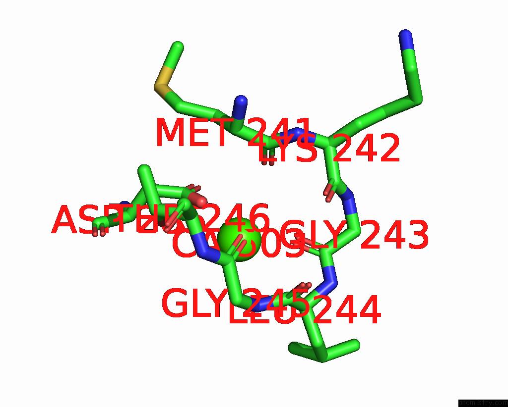
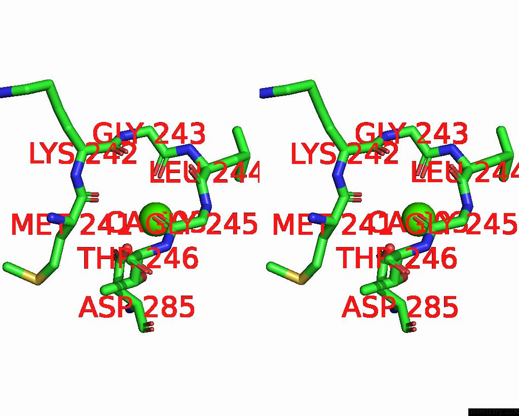
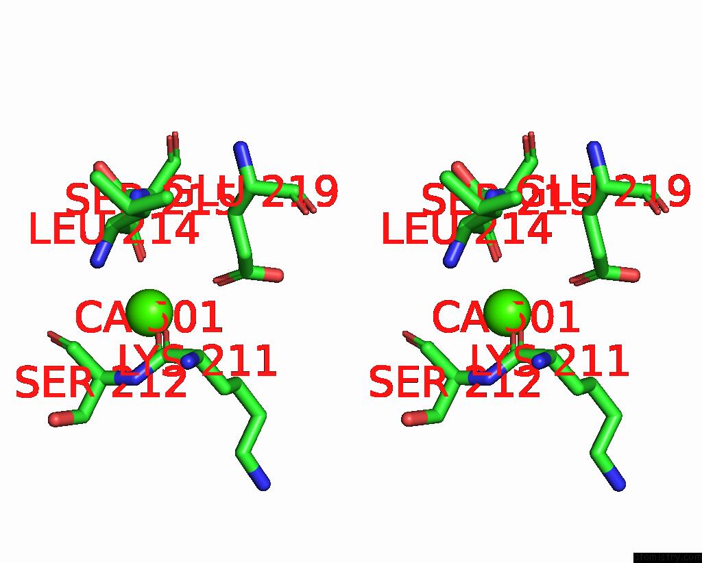
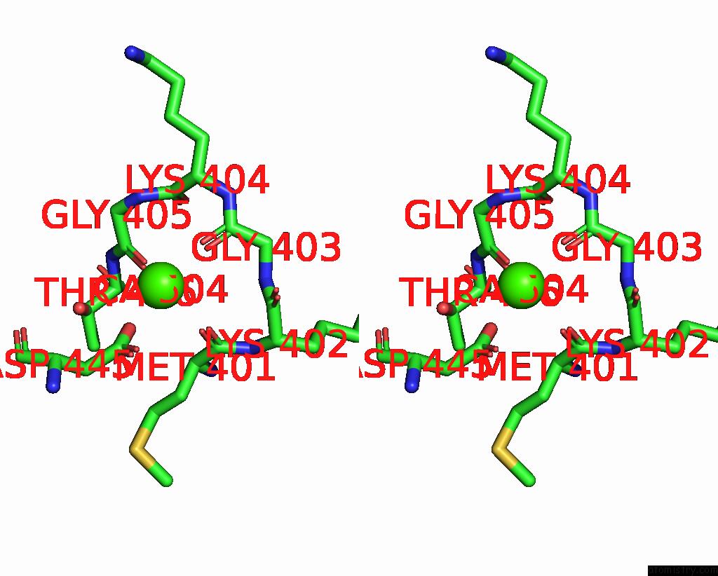
Calcium binding site 1 out of 9 in 7pc8
Go back to
Calcium binding site 1 out
of 9 in the The Pdz Domain of SNTG1 Complexed with the Phosphomimetic Mutant Pdz- Binding Motif of RSK1
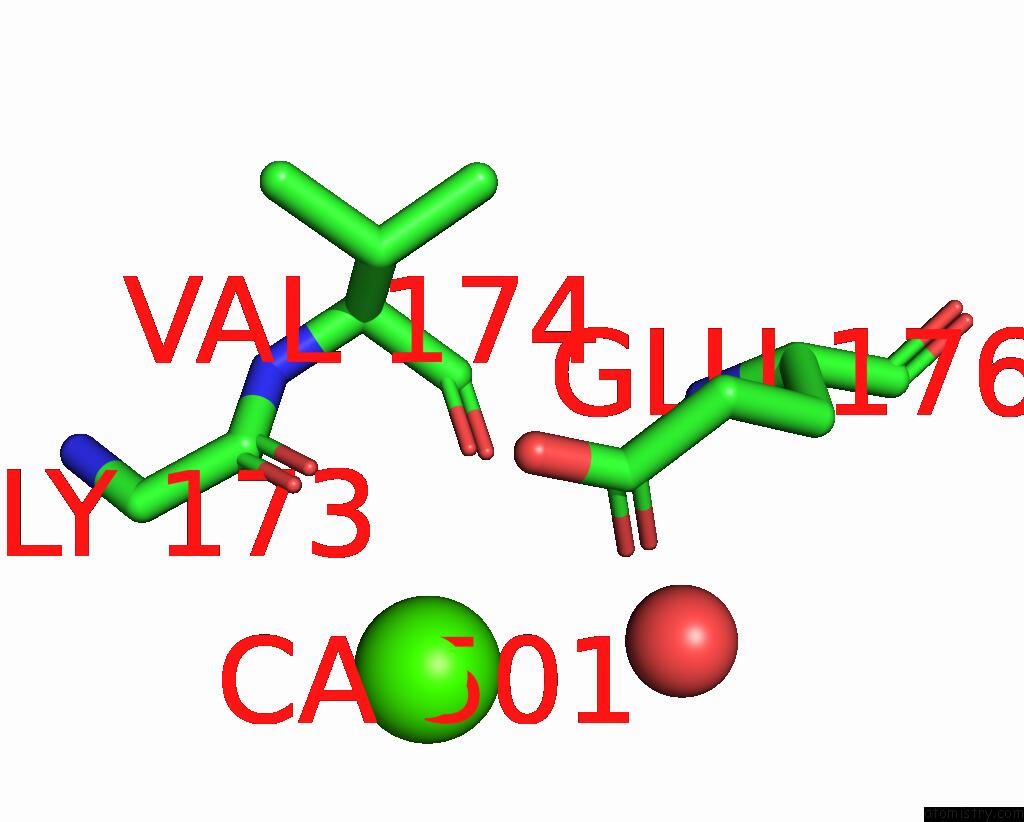
Mono view
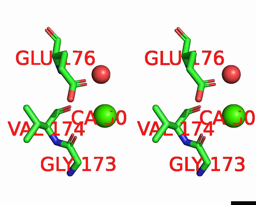
Stereo pair view
Mono view
Stereo pair view
A full contact list of Calcium with other atoms in the Ca binding
site number 1 of The Pdz Domain of SNTG1 Complexed with the Phosphomimetic Mutant Pdz- Binding Motif of RSK1 within 5.0Å range:
|
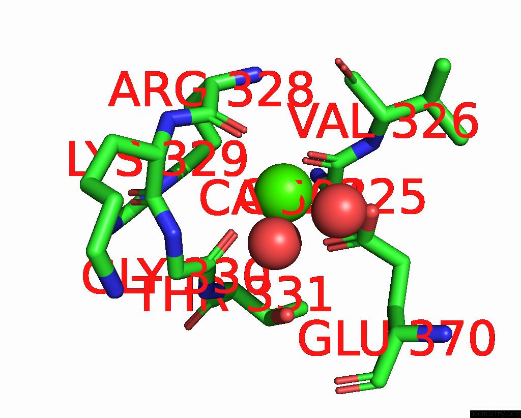
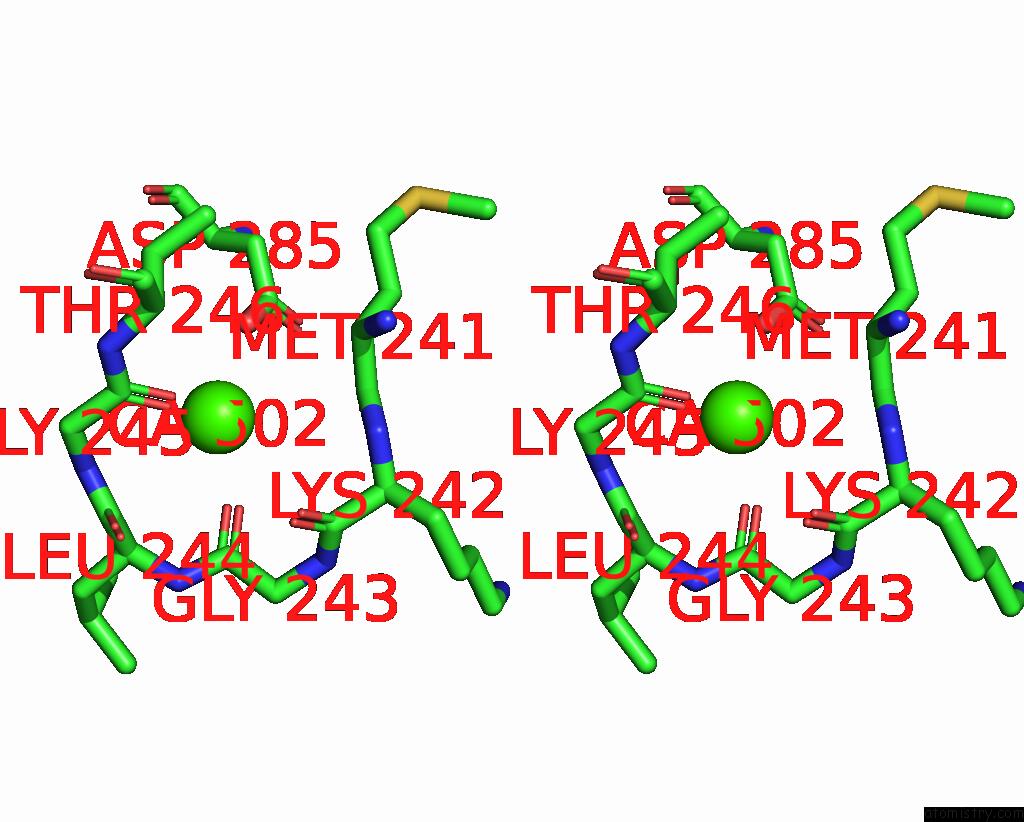
Calcium binding site 2 out of 9 in 7pc8
Go back to
Calcium binding site 2 out
of 9 in the The Pdz Domain of SNTG1 Complexed with the Phosphomimetic Mutant Pdz- Binding Motif of RSK1
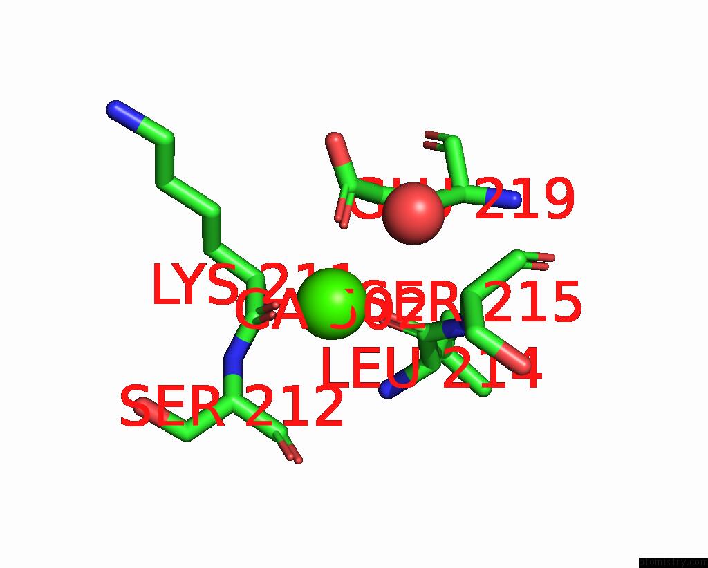
Mono view
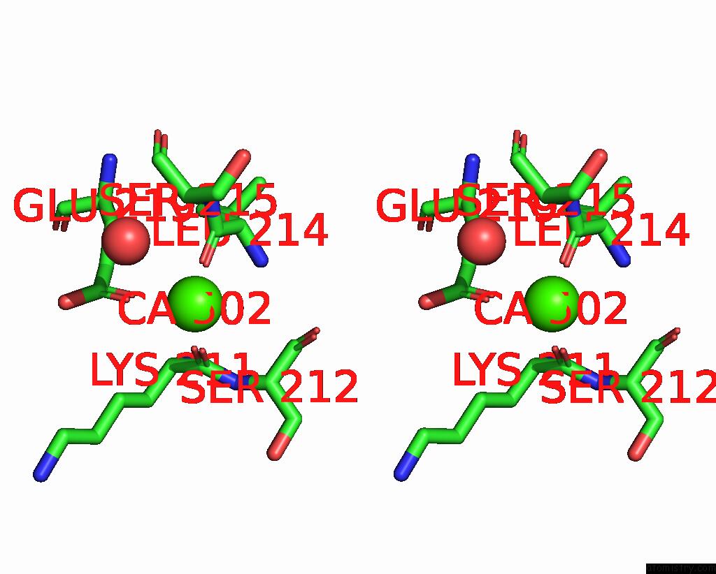
Stereo pair view
Mono view
Stereo pair view
A full contact list of Calcium with other atoms in the Ca binding
site number 2 of The Pdz Domain of SNTG1 Complexed with the Phosphomimetic Mutant Pdz- Binding Motif of RSK1 within 5.0Å range:
|
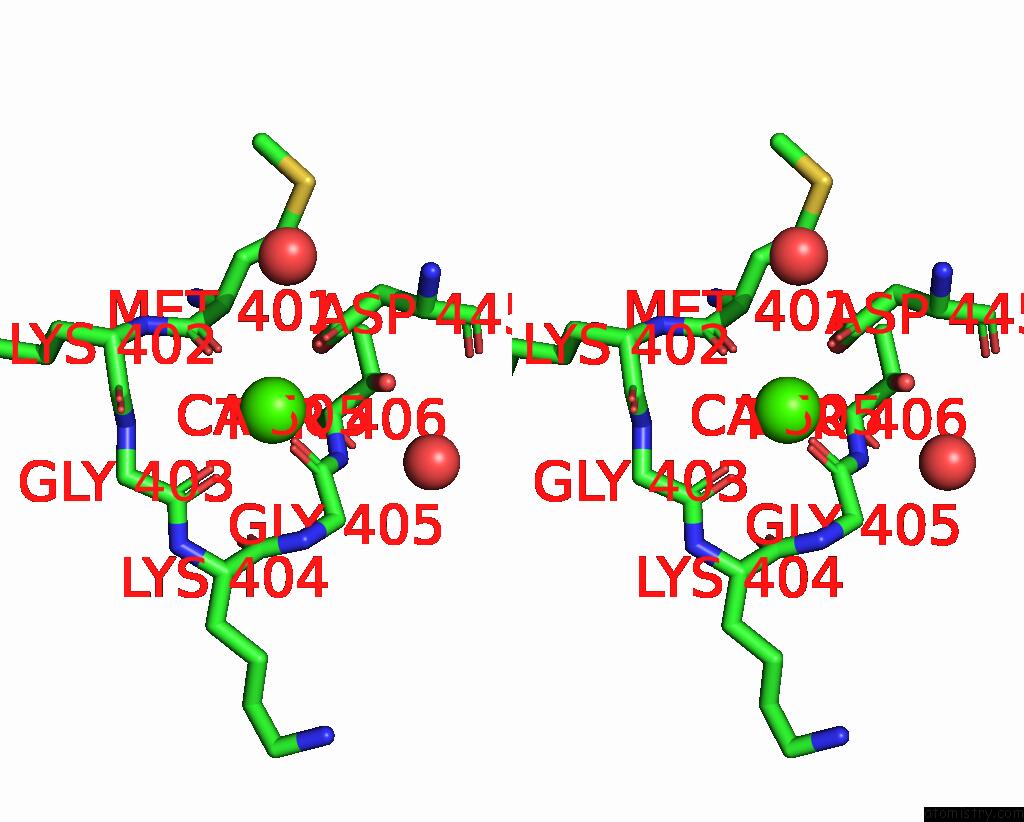
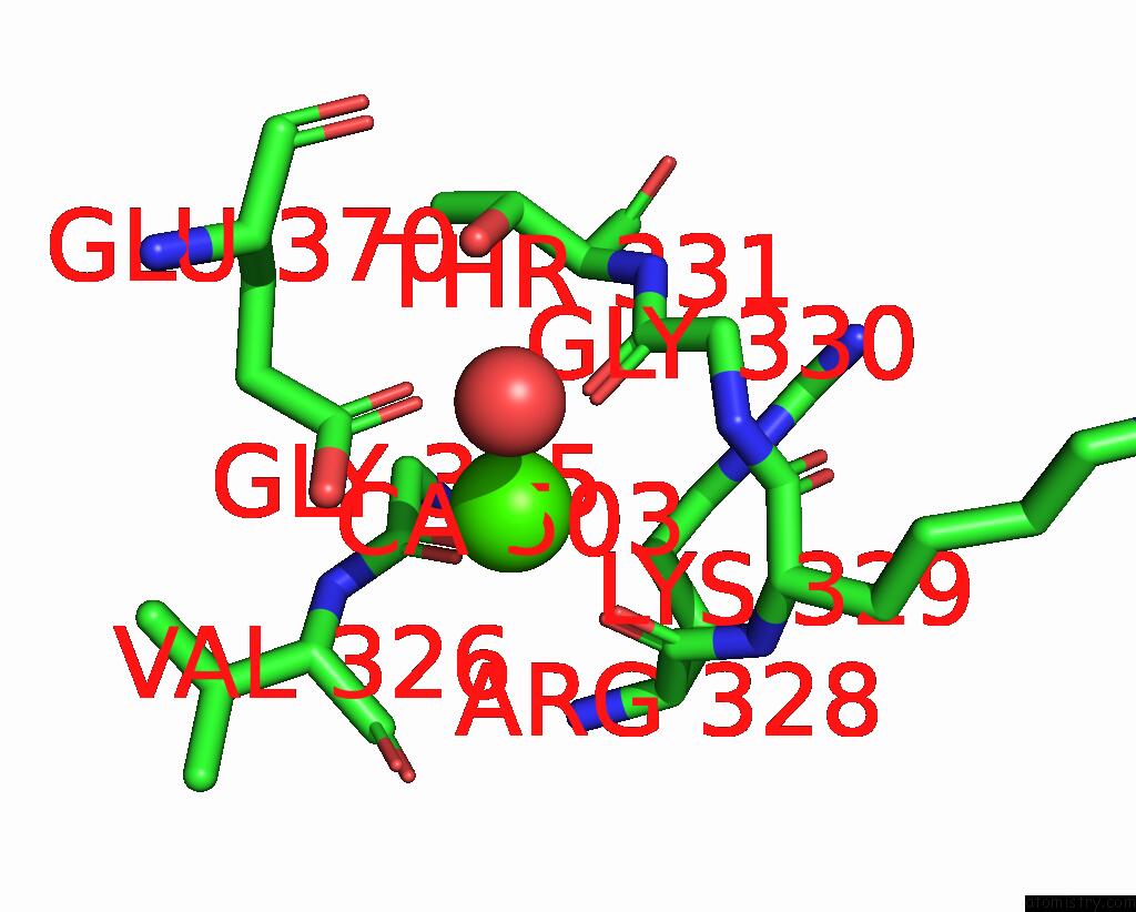
Calcium binding site 3 out of 9 in 7pc8
Go back to
Calcium binding site 3 out
of 9 in the The Pdz Domain of SNTG1 Complexed with the Phosphomimetic Mutant Pdz- Binding Motif of RSK1
Mono view
Stereo pair view
Mono view
Stereo pair view
A full contact list of Calcium with other atoms in the Ca binding
site number 3 of The Pdz Domain of SNTG1 Complexed with the Phosphomimetic Mutant Pdz- Binding Motif of RSK1 within 5.0Å range:
|
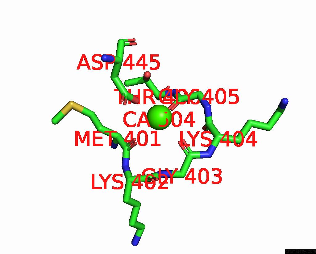
Calcium binding site 4 out of 9 in 7pc8
Go back to
Calcium binding site 4 out
of 9 in the The Pdz Domain of SNTG1 Complexed with the Phosphomimetic Mutant Pdz- Binding Motif of RSK1
Mono view

Stereo pair view
Mono view
Stereo pair view
A full contact list of Calcium with other atoms in the Ca binding
site number 4 of The Pdz Domain of SNTG1 Complexed with the Phosphomimetic Mutant Pdz- Binding Motif of RSK1 within 5.0Å range:
|
Calcium binding site 5 out of 9 in 7pc8
Go back to
Calcium binding site 5 out
of 9 in the The Pdz Domain of SNTG1 Complexed with the Phosphomimetic Mutant Pdz- Binding Motif of RSK1

Mono view
Stereo pair view
Mono view
Stereo pair view
A full contact list of Calcium with other atoms in the Ca binding
site number 5 of The Pdz Domain of SNTG1 Complexed with the Phosphomimetic Mutant Pdz- Binding Motif of RSK1 within 5.0Å range:
|
Calcium binding site 6 out of 9 in 7pc8
Go back to
Calcium binding site 6 out
of 9 in the The Pdz Domain of SNTG1 Complexed with the Phosphomimetic Mutant Pdz- Binding Motif of RSK1

Mono view
Stereo pair view
Mono view
Stereo pair view
A full contact list of Calcium with other atoms in the Ca binding
site number 6 of The Pdz Domain of SNTG1 Complexed with the Phosphomimetic Mutant Pdz- Binding Motif of RSK1 within 5.0Å range:
|
Calcium binding site 7 out of 9 in 7pc8
Go back to
Calcium binding site 7 out
of 9 in the The Pdz Domain of SNTG1 Complexed with the Phosphomimetic Mutant Pdz- Binding Motif of RSK1

Mono view
Stereo pair view
Mono view
Stereo pair view
A full contact list of Calcium with other atoms in the Ca binding
site number 7 of The Pdz Domain of SNTG1 Complexed with the Phosphomimetic Mutant Pdz- Binding Motif of RSK1 within 5.0Å range:
|
Calcium binding site 8 out of 9 in 7pc8
Go back to
Calcium binding site 8 out
of 9 in the The Pdz Domain of SNTG1 Complexed with the Phosphomimetic Mutant Pdz- Binding Motif of RSK1
Mono view

Stereo pair view
Mono view
Stereo pair view
A full contact list of Calcium with other atoms in the Ca binding
site number 8 of The Pdz Domain of SNTG1 Complexed with the Phosphomimetic Mutant Pdz- Binding Motif of RSK1 within 5.0Å range:
|
Calcium binding site 9 out of 9 in 7pc8
Go back to
Calcium binding site 9 out
of 9 in the The Pdz Domain of SNTG1 Complexed with the Phosphomimetic Mutant Pdz- Binding Motif of RSK1
Mono view
Stereo pair view
Mono view
Stereo pair view
A full contact list of Calcium with other atoms in the Ca binding
site number 9 of The Pdz Domain of SNTG1 Complexed with the Phosphomimetic Mutant Pdz- Binding Motif of RSK1 within 5.0Å range:
|
Reference:
A.Cousido-Siah,
L.Carneiro,
C.Kostmann,
P.Ecsedi,
L.Nyitray,
G.Trave,
G.Gogl.
A Scalable Strategy to Solve Structures of Pdz Domains and Their Complexes. Acta Crystallogr D Struct V. 78 509 2022BIOL.
ISSN: ISSN 2059-7983
PubMed: 35362473
DOI: 10.1107/S2059798322001784
Page generated: Thu Jul 10 00:06:39 2025
ISSN: ISSN 2059-7983
PubMed: 35362473
DOI: 10.1107/S2059798322001784
Last articles
Fe in 2YXOFe in 2YRS
Fe in 2YXC
Fe in 2YNM
Fe in 2YVJ
Fe in 2YP1
Fe in 2YU2
Fe in 2YU1
Fe in 2YQB
Fe in 2YOO