Calcium »
PDB 7p9t-7po7 »
7pjm »
Calcium in PDB 7pjm: Crystal Structure of Ivosidenib-Resistant IDH1 Variant R132C S280F in Complex with Nadph and CA2+/2-Oxoglutarate
Enzymatic activity of Crystal Structure of Ivosidenib-Resistant IDH1 Variant R132C S280F in Complex with Nadph and CA2+/2-Oxoglutarate
All present enzymatic activity of Crystal Structure of Ivosidenib-Resistant IDH1 Variant R132C S280F in Complex with Nadph and CA2+/2-Oxoglutarate:
1.1.1.42;
1.1.1.42;
Protein crystallography data
The structure of Crystal Structure of Ivosidenib-Resistant IDH1 Variant R132C S280F in Complex with Nadph and CA2+/2-Oxoglutarate, PDB code: 7pjm
was solved by
R.Reinbold,
P.Rabe,
M.I.Abboud,
C.J.Schofield,
with X-Ray Crystallography technique. A brief refinement statistics is given in the table below:
| Resolution Low / High (Å) | 59.01 / 2.10 |
| Space group | C 2 2 21 |
| Cell size a, b, c (Å), α, β, γ (°) | 96.58, 273.01, 117.42, 90, 90, 90 |
| R / Rfree (%) | 18.1 / 21 |
Other elements in 7pjm:
The structure of Crystal Structure of Ivosidenib-Resistant IDH1 Variant R132C S280F in Complex with Nadph and CA2+/2-Oxoglutarate also contains other interesting chemical elements:
| Chlorine | (Cl) | 3 atoms |
Calcium Binding Sites:
The binding sites of Calcium atom in the Crystal Structure of Ivosidenib-Resistant IDH1 Variant R132C S280F in Complex with Nadph and CA2+/2-Oxoglutarate
(pdb code 7pjm). This binding sites where shown within
5.0 Angstroms radius around Calcium atom.
In total 3 binding sites of Calcium where determined in the Crystal Structure of Ivosidenib-Resistant IDH1 Variant R132C S280F in Complex with Nadph and CA2+/2-Oxoglutarate, PDB code: 7pjm:
Jump to Calcium binding site number: 1; 2; 3;
In total 3 binding sites of Calcium where determined in the Crystal Structure of Ivosidenib-Resistant IDH1 Variant R132C S280F in Complex with Nadph and CA2+/2-Oxoglutarate, PDB code: 7pjm:
Jump to Calcium binding site number: 1; 2; 3;
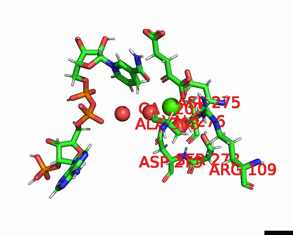
Calcium binding site 1 out of 3 in 7pjm
Go back to
Calcium binding site 1 out
of 3 in the Crystal Structure of Ivosidenib-Resistant IDH1 Variant R132C S280F in Complex with Nadph and CA2+/2-Oxoglutarate
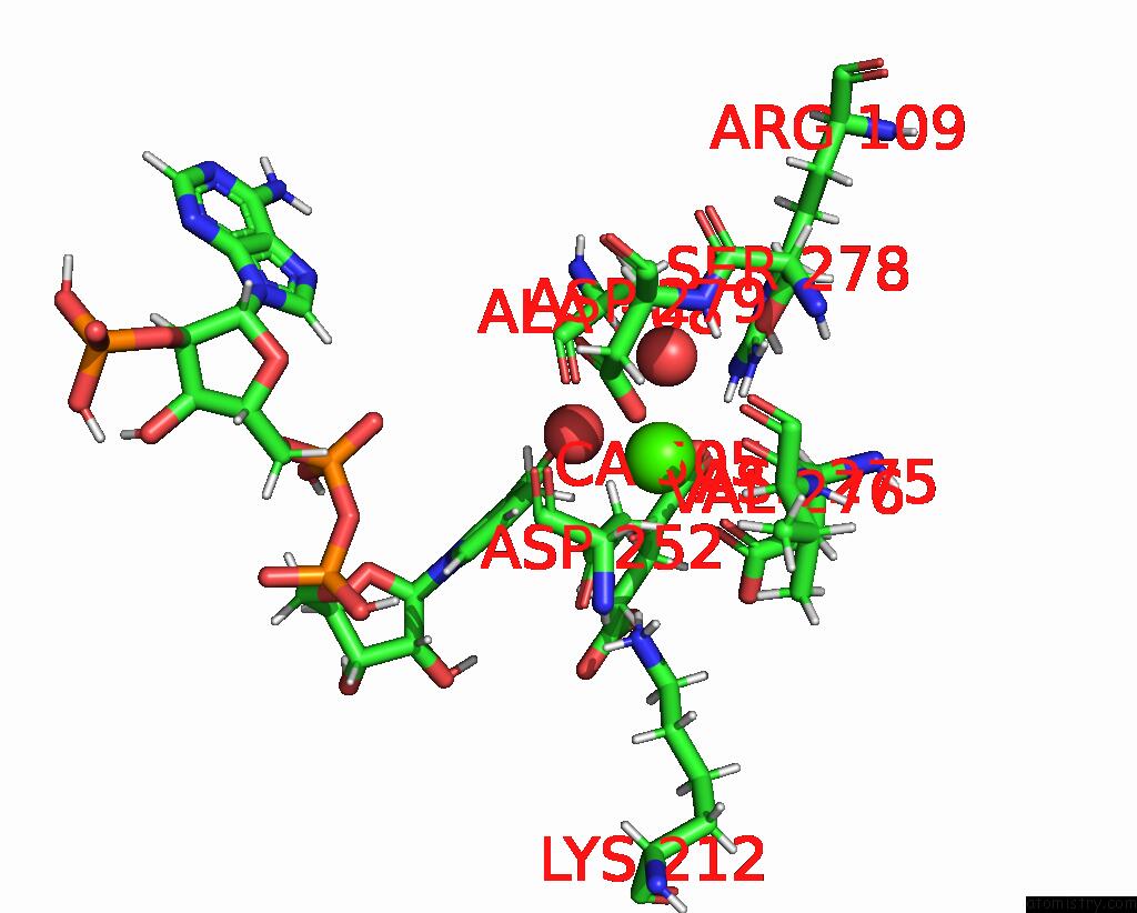
Mono view
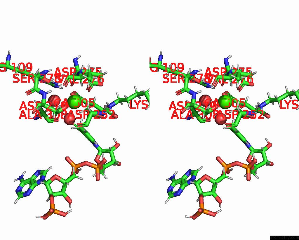
Stereo pair view
Mono view
Stereo pair view
A full contact list of Calcium with other atoms in the Ca binding
site number 1 of Crystal Structure of Ivosidenib-Resistant IDH1 Variant R132C S280F in Complex with Nadph and CA2+/2-Oxoglutarate within 5.0Å range:
|
Calcium binding site 2 out of 3 in 7pjm
Go back to
Calcium binding site 2 out
of 3 in the Crystal Structure of Ivosidenib-Resistant IDH1 Variant R132C S280F in Complex with Nadph and CA2+/2-Oxoglutarate
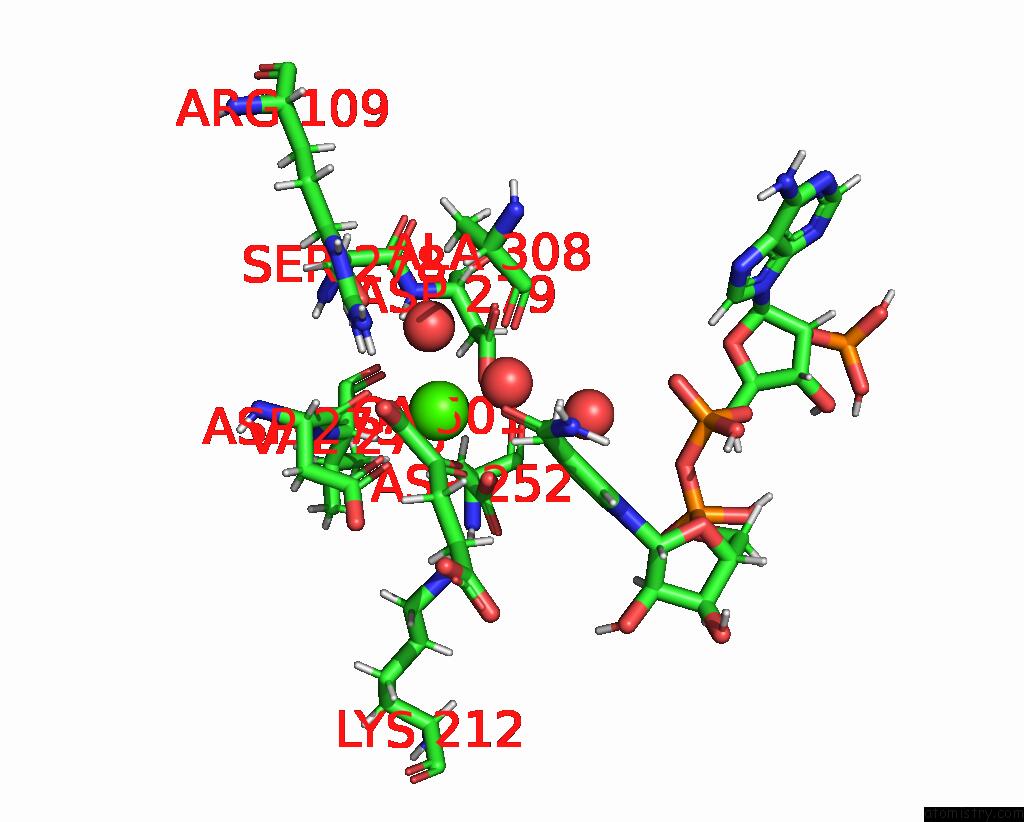
Mono view
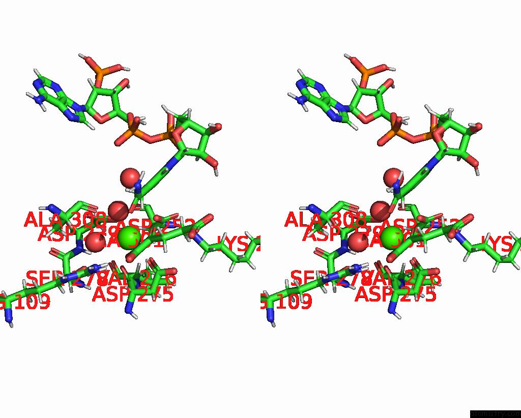
Stereo pair view
Mono view
Stereo pair view
A full contact list of Calcium with other atoms in the Ca binding
site number 2 of Crystal Structure of Ivosidenib-Resistant IDH1 Variant R132C S280F in Complex with Nadph and CA2+/2-Oxoglutarate within 5.0Å range:
|
Calcium binding site 3 out of 3 in 7pjm
Go back to
Calcium binding site 3 out
of 3 in the Crystal Structure of Ivosidenib-Resistant IDH1 Variant R132C S280F in Complex with Nadph and CA2+/2-Oxoglutarate
Mono view

Stereo pair view
Mono view
Stereo pair view
A full contact list of Calcium with other atoms in the Ca binding
site number 3 of Crystal Structure of Ivosidenib-Resistant IDH1 Variant R132C S280F in Complex with Nadph and CA2+/2-Oxoglutarate within 5.0Å range:
|
Reference:
R.Reinbold,
I.C.Hvinden,
P.Rabe,
R.A.Herold,
A.Finch,
J.Wood,
M.Morgan,
M.Staudt,
I.J.Clifton,
F.A.Armstrong,
J.S.O.Mccullagh,
J.Redmond,
C.Bardella,
M.I.Abboud,
C.J.Schofield.
Resistance to the Isocitrate Dehydrogenase 1 Mutant Inhibitor Ivosidenib Can Be Overcome By Alternative Dimer-Interface Binding Inhibitors. Nat Commun V. 13 4785 2022.
ISSN: ESSN 2041-1723
PubMed: 35970853
DOI: 10.1038/S41467-022-32436-4
Page generated: Thu Jul 10 00:09:51 2025
ISSN: ESSN 2041-1723
PubMed: 35970853
DOI: 10.1038/S41467-022-32436-4
Last articles
Cl in 5L4RCl in 5L55
Cl in 5L5A
Cl in 5L4M
Cl in 5L54
Cl in 5L52
Cl in 5L4H
Cl in 5L4E
Cl in 5L4I
Cl in 5L47