Calcium »
PDB 7qga-7rcc »
7qql »
Calcium in PDB 7qql: The Pdz Domain of SNTG2 Complexed with the Phosphorylated Pdz-Binding Motif of RSK1
Enzymatic activity of The Pdz Domain of SNTG2 Complexed with the Phosphorylated Pdz-Binding Motif of RSK1
All present enzymatic activity of The Pdz Domain of SNTG2 Complexed with the Phosphorylated Pdz-Binding Motif of RSK1:
2.7.11.1;
2.7.11.1;
Protein crystallography data
The structure of The Pdz Domain of SNTG2 Complexed with the Phosphorylated Pdz-Binding Motif of RSK1, PDB code: 7qql
was solved by
A.Cousido-Siah,
G.Trave,
G.Gogl,
with X-Ray Crystallography technique. A brief refinement statistics is given in the table below:
| Resolution Low / High (Å) | 48.42 / 2.44 |
| Space group | P 1 21 1 |
| Cell size a, b, c (Å), α, β, γ (°) | 97.46, 60.55, 135.21, 90, 92.42, 90 |
| R / Rfree (%) | 19.5 / 23 |
Calcium Binding Sites:
Pages:
>>> Page 1 <<< Page 2, Binding sites: 11 - 14;Binding sites:
The binding sites of Calcium atom in the The Pdz Domain of SNTG2 Complexed with the Phosphorylated Pdz-Binding Motif of RSK1 (pdb code 7qql). This binding sites where shown within 5.0 Angstroms radius around Calcium atom.In total 14 binding sites of Calcium where determined in the The Pdz Domain of SNTG2 Complexed with the Phosphorylated Pdz-Binding Motif of RSK1, PDB code: 7qql:
Jump to Calcium binding site number: 1; 2; 3; 4; 5; 6; 7; 8; 9; 10;
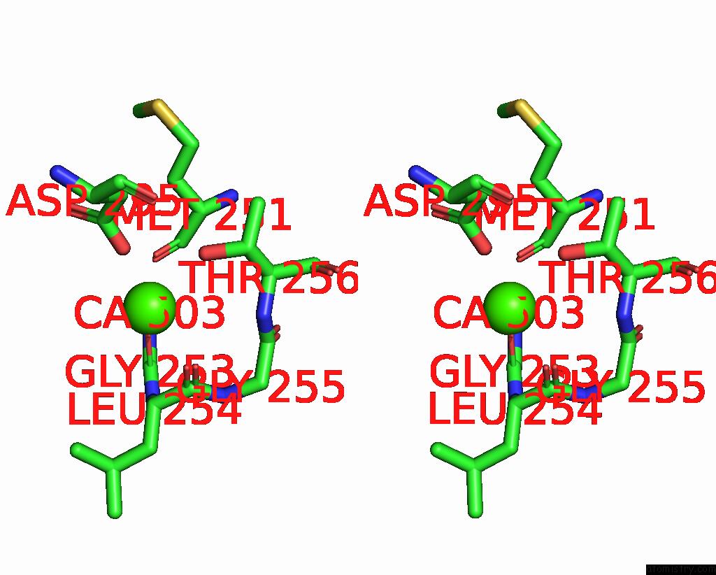
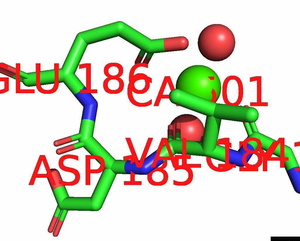
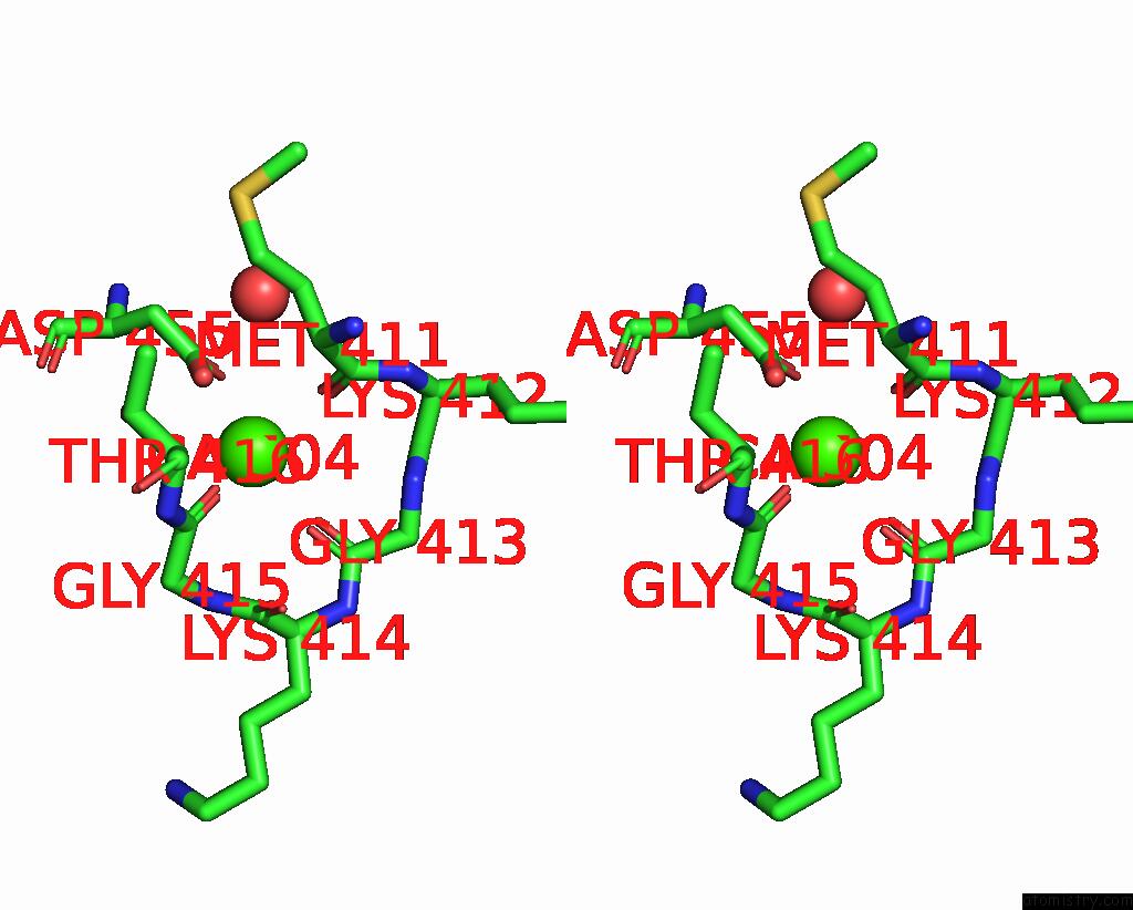
Calcium binding site 1 out of 14 in 7qql
Go back to
Calcium binding site 1 out
of 14 in the The Pdz Domain of SNTG2 Complexed with the Phosphorylated Pdz-Binding Motif of RSK1
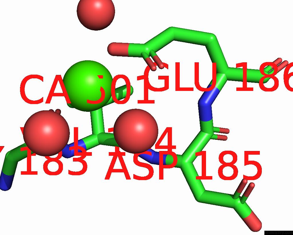
Mono view
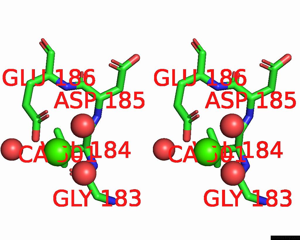
Stereo pair view
Mono view
Stereo pair view
|
|
A full contact list of Calcium with other atoms in the Ca binding
site number 1 of The Pdz Domain of SNTG2 Complexed with the Phosphorylated Pdz-Binding Motif of RSK1 within 5.0Å range:
|
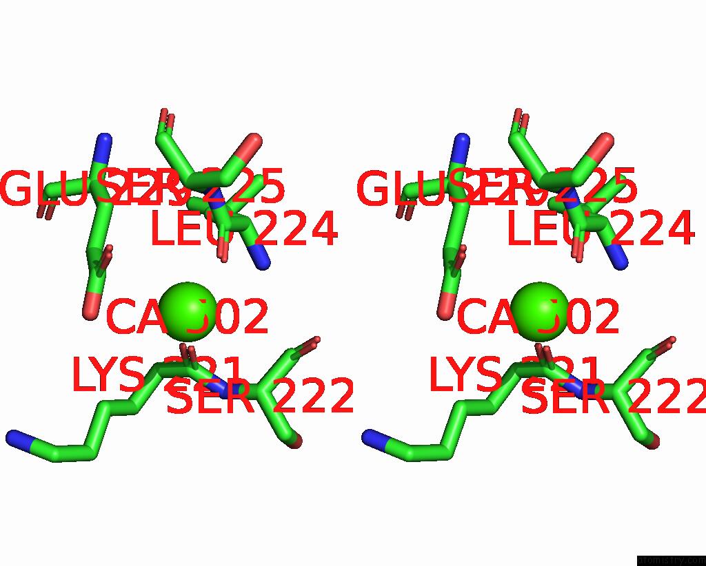
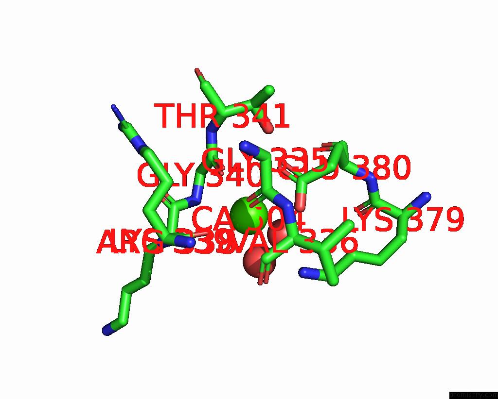
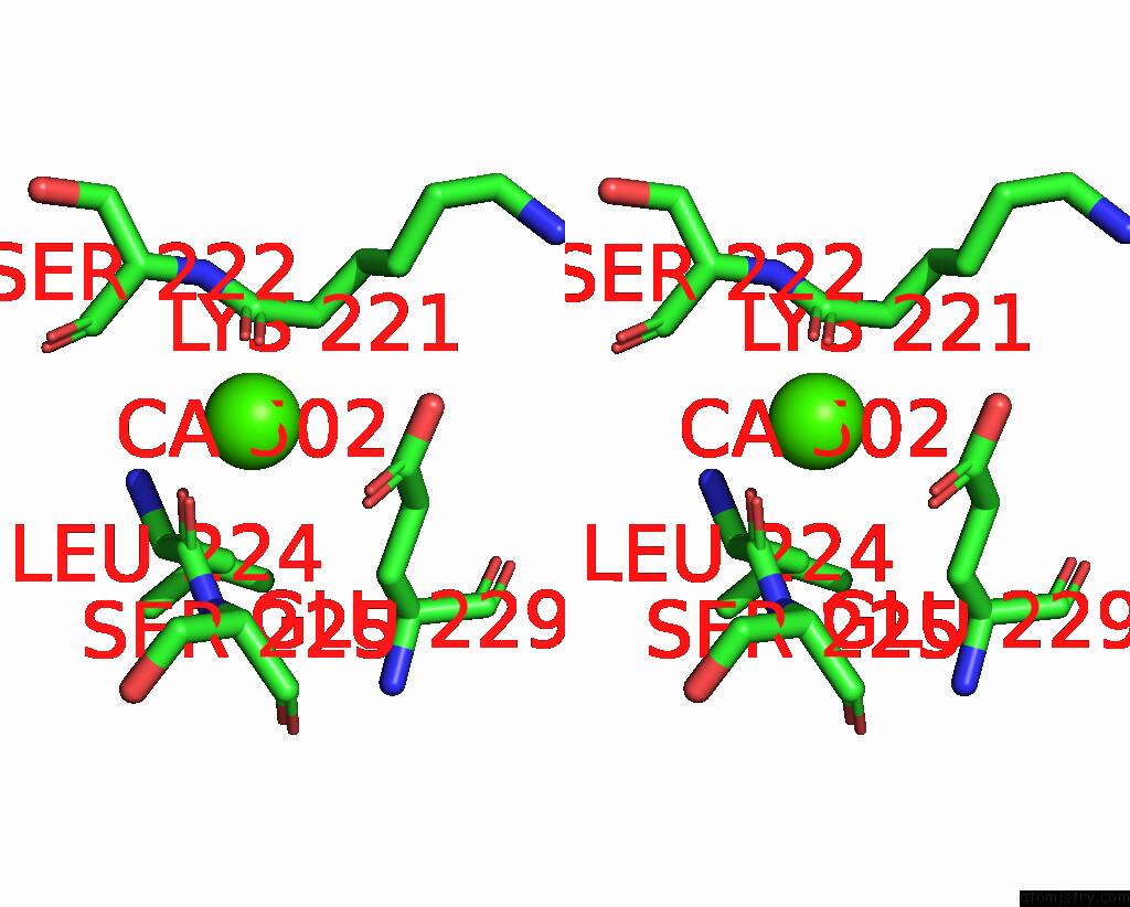
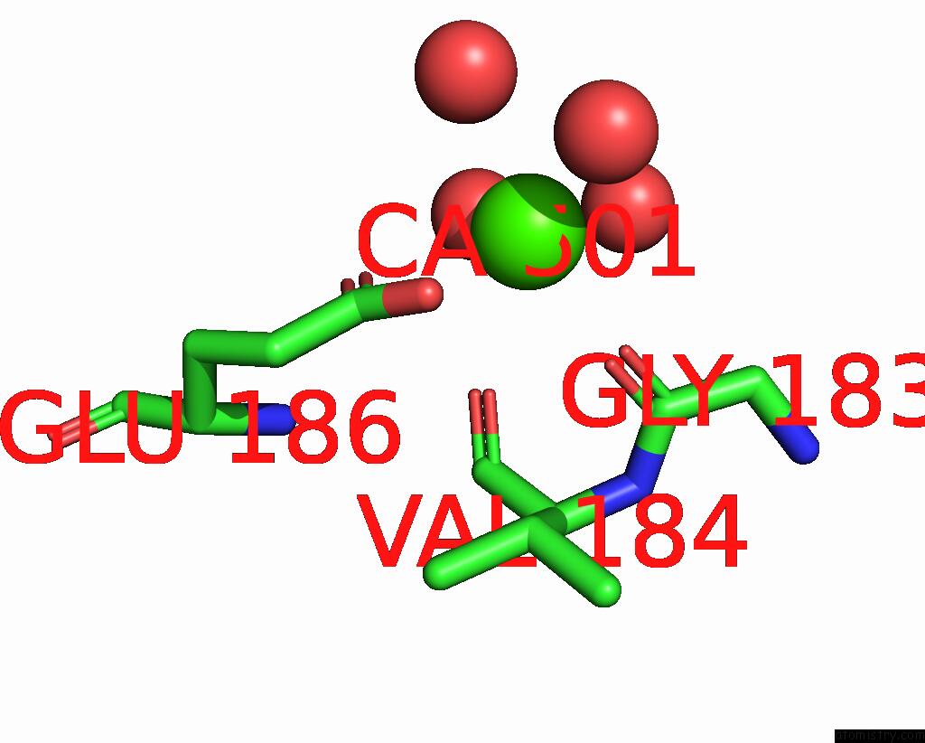
Calcium binding site 2 out of 14 in 7qql
Go back to
Calcium binding site 2 out
of 14 in the The Pdz Domain of SNTG2 Complexed with the Phosphorylated Pdz-Binding Motif of RSK1
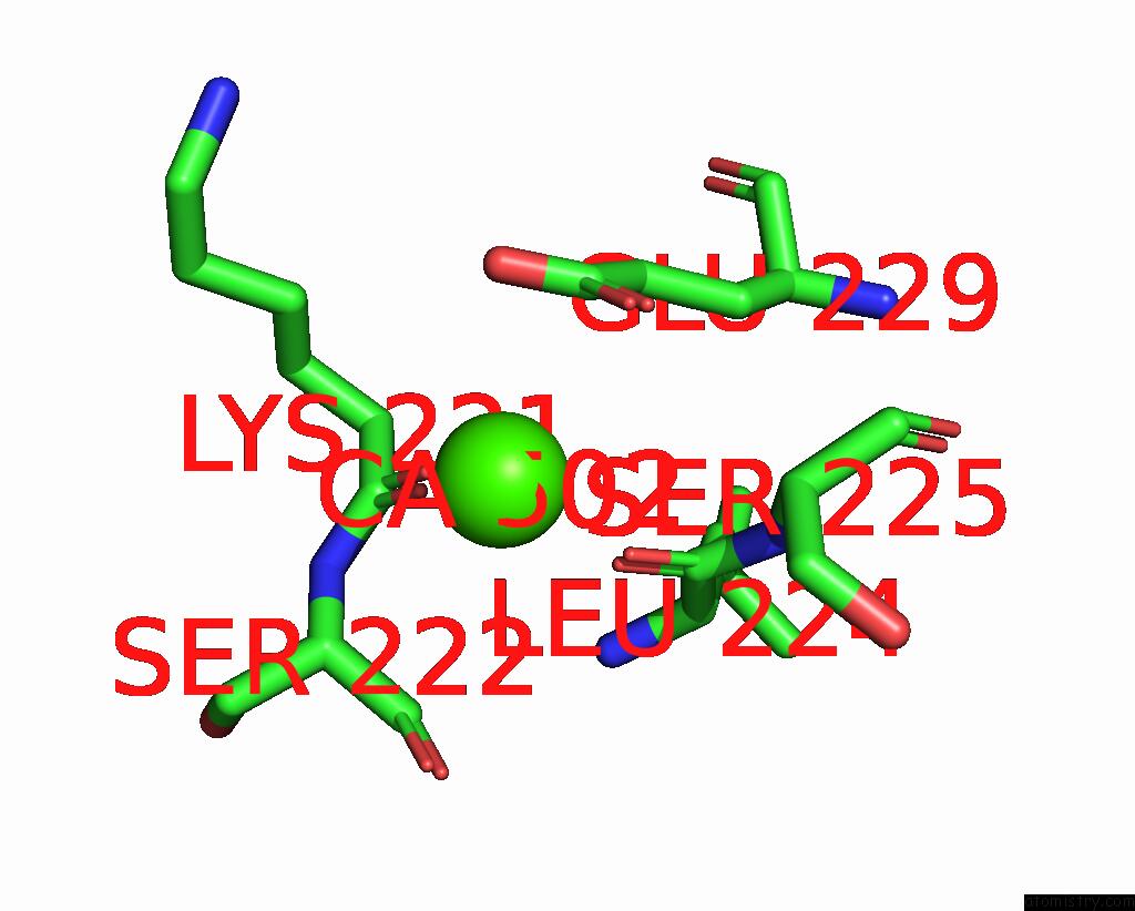
Mono view
Stereo pair view
Mono view
Stereo pair view
|
|
A full contact list of Calcium with other atoms in the Ca binding
site number 2 of The Pdz Domain of SNTG2 Complexed with the Phosphorylated Pdz-Binding Motif of RSK1 within 5.0Å range:
|
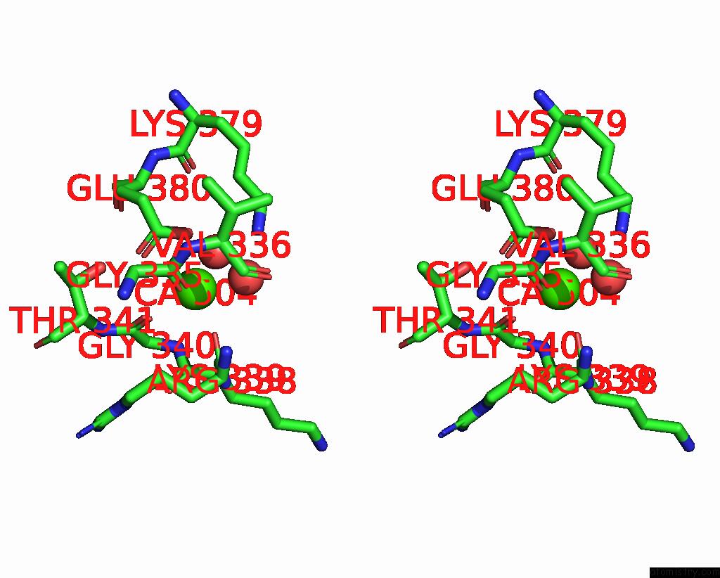
Calcium binding site 3 out of 14 in 7qql
Go back to
Calcium binding site 3 out
of 14 in the The Pdz Domain of SNTG2 Complexed with the Phosphorylated Pdz-Binding Motif of RSK1

Mono view
Stereo pair view
Mono view
Stereo pair view
|
|
A full contact list of Calcium with other atoms in the Ca binding
site number 3 of The Pdz Domain of SNTG2 Complexed with the Phosphorylated Pdz-Binding Motif of RSK1 within 5.0Å range:
|
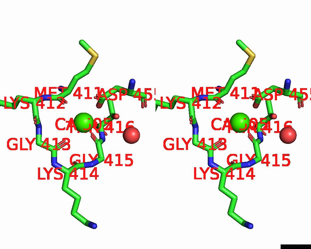
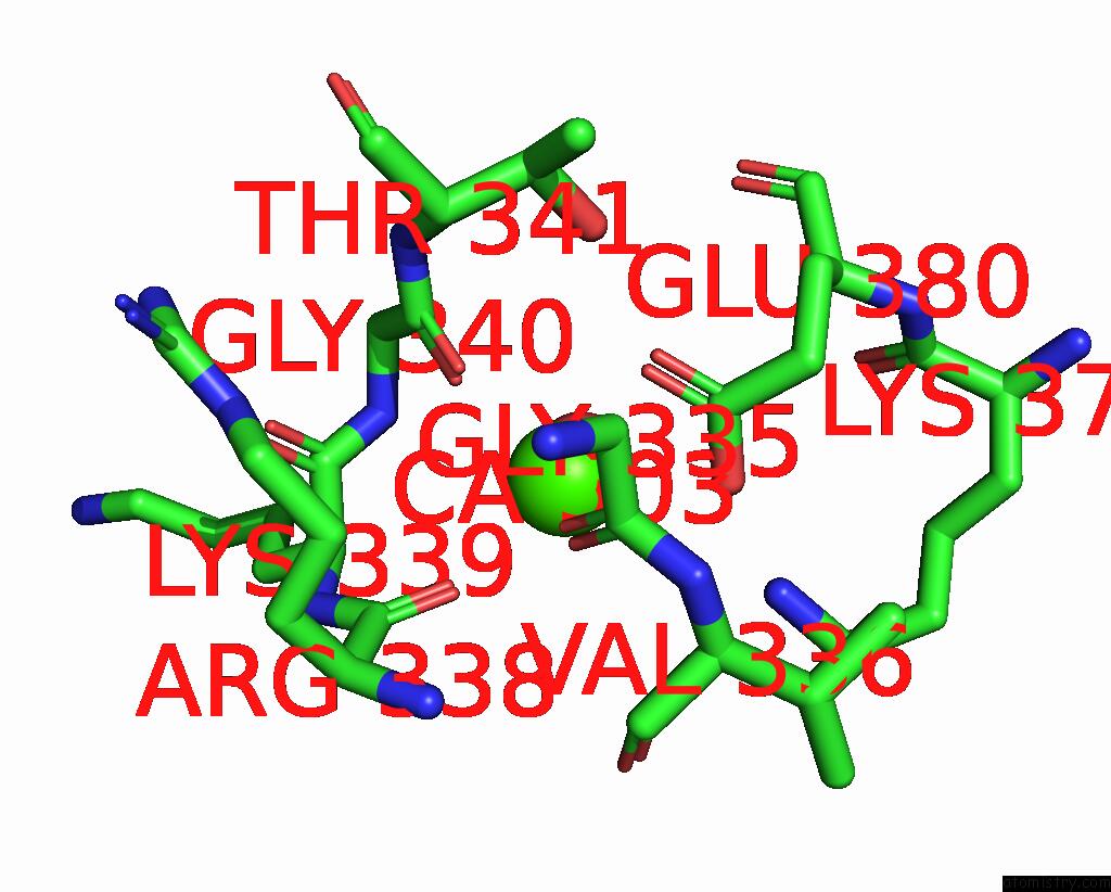
Calcium binding site 4 out of 14 in 7qql
Go back to
Calcium binding site 4 out
of 14 in the The Pdz Domain of SNTG2 Complexed with the Phosphorylated Pdz-Binding Motif of RSK1
Mono view
Stereo pair view
Mono view
Stereo pair view
|
|
A full contact list of Calcium with other atoms in the Ca binding
site number 4 of The Pdz Domain of SNTG2 Complexed with the Phosphorylated Pdz-Binding Motif of RSK1 within 5.0Å range:
|
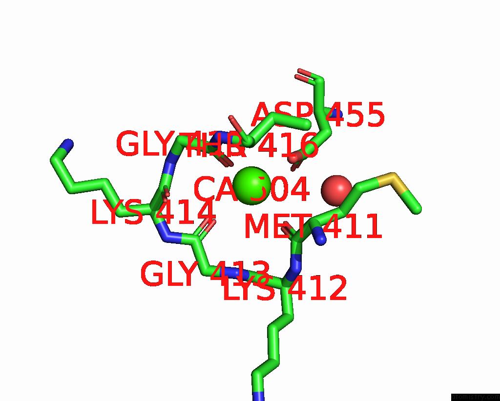
Calcium binding site 5 out of 14 in 7qql
Go back to
Calcium binding site 5 out
of 14 in the The Pdz Domain of SNTG2 Complexed with the Phosphorylated Pdz-Binding Motif of RSK1

Mono view
Stereo pair view
Mono view
Stereo pair view
|
|
A full contact list of Calcium with other atoms in the Ca binding
site number 5 of The Pdz Domain of SNTG2 Complexed with the Phosphorylated Pdz-Binding Motif of RSK1 within 5.0Å range:
|
Calcium binding site 6 out of 14 in 7qql
Go back to
Calcium binding site 6 out
of 14 in the The Pdz Domain of SNTG2 Complexed with the Phosphorylated Pdz-Binding Motif of RSK1
Mono view

Stereo pair view
Mono view
Stereo pair view
|
|
A full contact list of Calcium with other atoms in the Ca binding
site number 6 of The Pdz Domain of SNTG2 Complexed with the Phosphorylated Pdz-Binding Motif of RSK1 within 5.0Å range:
|
Calcium binding site 7 out of 14 in 7qql
Go back to
Calcium binding site 7 out
of 14 in the The Pdz Domain of SNTG2 Complexed with the Phosphorylated Pdz-Binding Motif of RSK1

Mono view
Stereo pair view
Mono view
Stereo pair view
|
|
A full contact list of Calcium with other atoms in the Ca binding
site number 7 of The Pdz Domain of SNTG2 Complexed with the Phosphorylated Pdz-Binding Motif of RSK1 within 5.0Å range:
|
Calcium binding site 8 out of 14 in 7qql
Go back to
Calcium binding site 8 out
of 14 in the The Pdz Domain of SNTG2 Complexed with the Phosphorylated Pdz-Binding Motif of RSK1
Mono view

Stereo pair view
Mono view
Stereo pair view
|
|
A full contact list of Calcium with other atoms in the Ca binding
site number 8 of The Pdz Domain of SNTG2 Complexed with the Phosphorylated Pdz-Binding Motif of RSK1 within 5.0Å range:
|
Calcium binding site 9 out of 14 in 7qql
Go back to
Calcium binding site 9 out
of 14 in the The Pdz Domain of SNTG2 Complexed with the Phosphorylated Pdz-Binding Motif of RSK1
Mono view
Stereo pair view
Mono view
Stereo pair view
|
|
A full contact list of Calcium with other atoms in the Ca binding
site number 9 of The Pdz Domain of SNTG2 Complexed with the Phosphorylated Pdz-Binding Motif of RSK1 within 5.0Å range:
|
Calcium binding site 10 out of 14 in 7qql
Go back to
Calcium binding site 10 out
of 14 in the The Pdz Domain of SNTG2 Complexed with the Phosphorylated Pdz-Binding Motif of RSK1
Mono view

Stereo pair view
Mono view
Stereo pair view
|
|
A full contact list of Calcium with other atoms in the Ca binding
site number 10 of The Pdz Domain of SNTG2 Complexed with the Phosphorylated Pdz-Binding Motif of RSK1 within 5.0Å range:
|
Reference:
A.Cousido-Siah,
L.Carneiro,
C.Kostmann,
P.Ecsedi,
L.Nyitray,
G.Trave,
G.Gogl.
A Scalable Strategy to Solve Structures of Pdz Domains and Their Complexes. Acta Crystallogr D Struct V. 78 509 2022BIOL.
ISSN: ISSN 2059-7983
PubMed: 35362473
DOI: 10.1107/S2059798322001784
Page generated: Fri Jul 19 03:30:04 2024
ISSN: ISSN 2059-7983
PubMed: 35362473
DOI: 10.1107/S2059798322001784
Last articles
Zn in 9JYWZn in 9IR4
Zn in 9IR3
Zn in 9GMX
Zn in 9GMW
Zn in 9JEJ
Zn in 9ERF
Zn in 9ERE
Zn in 9EGV
Zn in 9EGW