Calcium »
PDB 7qga-7rcc »
7r3y »
Calcium in PDB 7r3y: The Crystal Structure of the V426L Variant of POL2CORE in Complex with Dna and An Incoming Nucleotide
Enzymatic activity of The Crystal Structure of the V426L Variant of POL2CORE in Complex with Dna and An Incoming Nucleotide
All present enzymatic activity of The Crystal Structure of the V426L Variant of POL2CORE in Complex with Dna and An Incoming Nucleotide:
2.7.7.7;
2.7.7.7;
Protein crystallography data
The structure of The Crystal Structure of the V426L Variant of POL2CORE in Complex with Dna and An Incoming Nucleotide, PDB code: 7r3y
was solved by
S.R.Barbari,
A.K.Beach,
J.G.Markgren,
V.Parkash,
E.Johansson,
P.V.Shcherbakova,
with X-Ray Crystallography technique. A brief refinement statistics is given in the table below:
| Resolution Low / High (Å) | 47.84 / 2.60 |
| Space group | P 1 2 1 |
| Cell size a, b, c (Å), α, β, γ (°) | 154.4, 70.1, 159.5, 90, 113.1, 90 |
| R / Rfree (%) | 22 / 27 |
Calcium Binding Sites:
The binding sites of Calcium atom in the The Crystal Structure of the V426L Variant of POL2CORE in Complex with Dna and An Incoming Nucleotide
(pdb code 7r3y). This binding sites where shown within
5.0 Angstroms radius around Calcium atom.
In total 4 binding sites of Calcium where determined in the The Crystal Structure of the V426L Variant of POL2CORE in Complex with Dna and An Incoming Nucleotide, PDB code: 7r3y:
Jump to Calcium binding site number: 1; 2; 3; 4;
In total 4 binding sites of Calcium where determined in the The Crystal Structure of the V426L Variant of POL2CORE in Complex with Dna and An Incoming Nucleotide, PDB code: 7r3y:
Jump to Calcium binding site number: 1; 2; 3; 4;
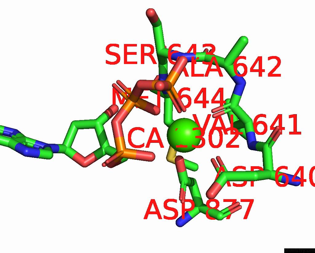
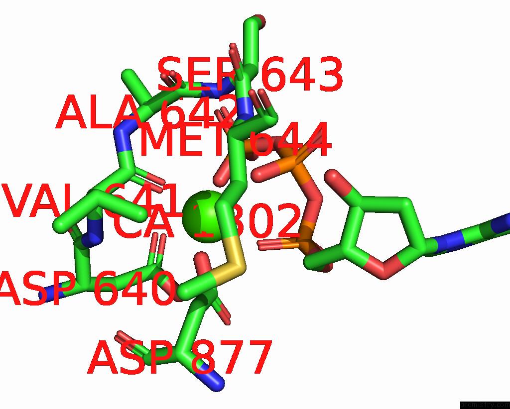
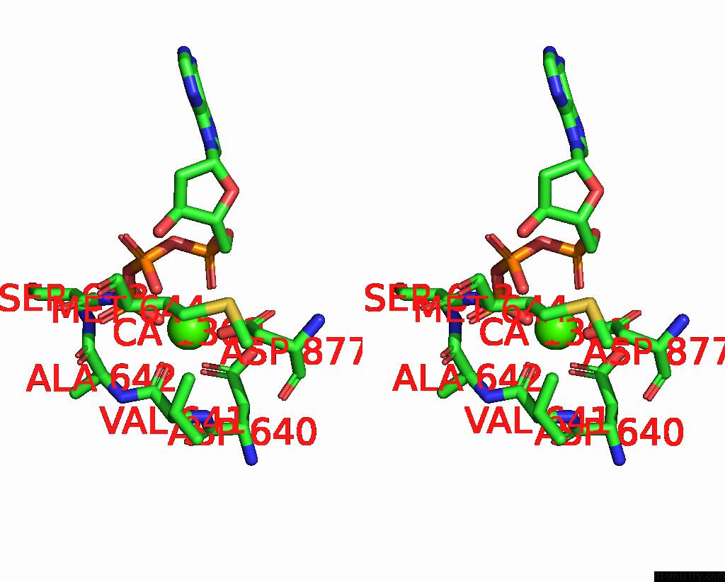
Calcium binding site 1 out of 4 in 7r3y
Go back to
Calcium binding site 1 out
of 4 in the The Crystal Structure of the V426L Variant of POL2CORE in Complex with Dna and An Incoming Nucleotide
Mono view
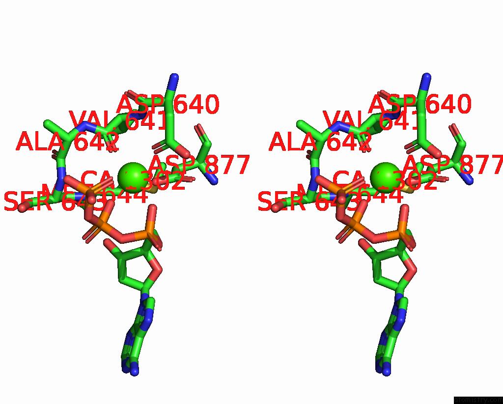
Stereo pair view
Mono view
Stereo pair view
A full contact list of Calcium with other atoms in the Ca binding
site number 1 of The Crystal Structure of the V426L Variant of POL2CORE in Complex with Dna and An Incoming Nucleotide within 5.0Å range:
|
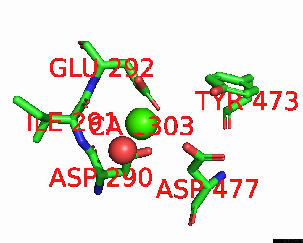
Calcium binding site 2 out of 4 in 7r3y
Go back to
Calcium binding site 2 out
of 4 in the The Crystal Structure of the V426L Variant of POL2CORE in Complex with Dna and An Incoming Nucleotide
Mono view
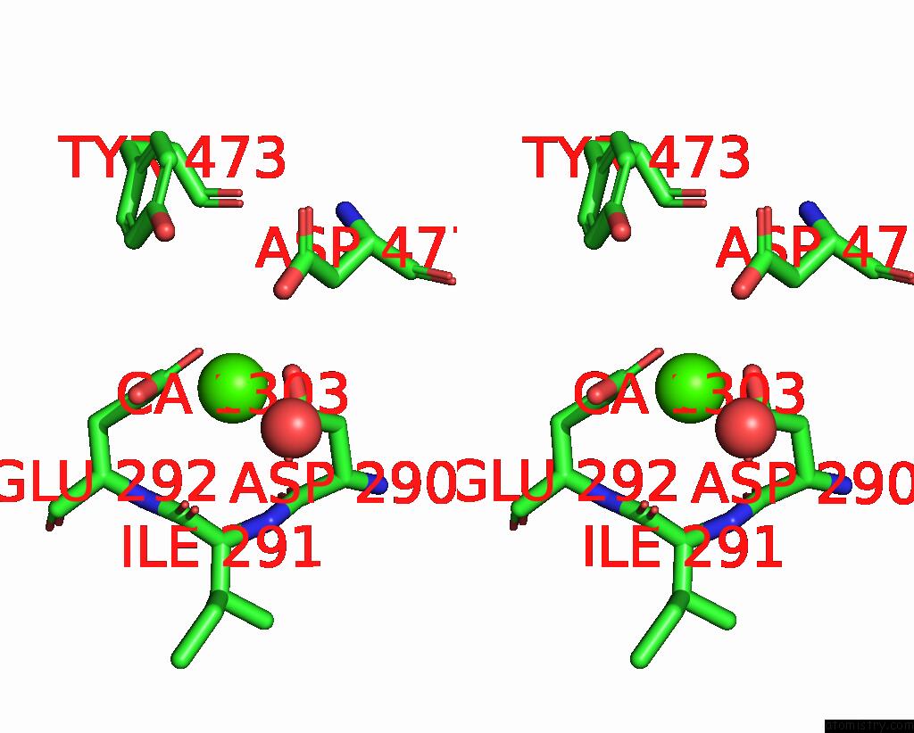
Stereo pair view
Mono view
Stereo pair view
A full contact list of Calcium with other atoms in the Ca binding
site number 2 of The Crystal Structure of the V426L Variant of POL2CORE in Complex with Dna and An Incoming Nucleotide within 5.0Å range:
|
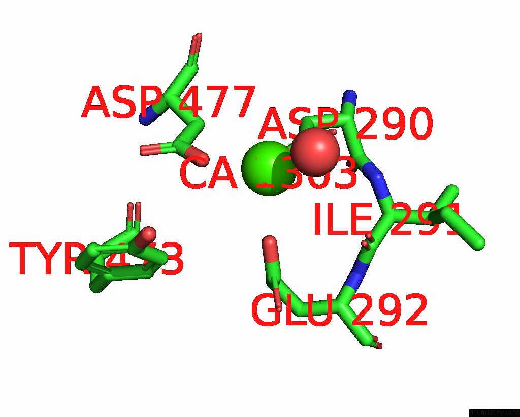
Calcium binding site 3 out of 4 in 7r3y
Go back to
Calcium binding site 3 out
of 4 in the The Crystal Structure of the V426L Variant of POL2CORE in Complex with Dna and An Incoming Nucleotide
Mono view
Stereo pair view
Mono view
Stereo pair view
A full contact list of Calcium with other atoms in the Ca binding
site number 3 of The Crystal Structure of the V426L Variant of POL2CORE in Complex with Dna and An Incoming Nucleotide within 5.0Å range:
|
Calcium binding site 4 out of 4 in 7r3y
Go back to
Calcium binding site 4 out
of 4 in the The Crystal Structure of the V426L Variant of POL2CORE in Complex with Dna and An Incoming Nucleotide
Mono view

Stereo pair view
Mono view
Stereo pair view
A full contact list of Calcium with other atoms in the Ca binding
site number 4 of The Crystal Structure of the V426L Variant of POL2CORE in Complex with Dna and An Incoming Nucleotide within 5.0Å range:
|
Reference:
S.R.Barbari,
A.K.Beach,
J.G.Markgren,
V.Parkash,
E.A.Moore,
E.Johansson,
P.V.Shcherbakova.
Enhanced Polymerase Activity Permits Efficient Synthesis By Cancer-Associated Dna Polymerase ε Variants at Low Dntp Levels. Nucleic Acids Res. V. 50 8023 2022.
ISSN: ESSN 1362-4962
PubMed: 35822874
DOI: 10.1093/NAR/GKAC602
Page generated: Thu Jul 10 00:41:10 2025
ISSN: ESSN 1362-4962
PubMed: 35822874
DOI: 10.1093/NAR/GKAC602
Last articles
Cl in 8B6PCl in 8B6R
Cl in 8B6Q
Cl in 8B6I
Cl in 8B6O
Cl in 8B5X
Cl in 8B5P
Cl in 8B2Y
Cl in 8B4X
Cl in 8B4W