Calcium »
PDB 7qga-7rcc »
7r4j »
Calcium in PDB 7r4j: Crystal Structure of Human Mitochondrial Nad Kinase
Enzymatic activity of Crystal Structure of Human Mitochondrial Nad Kinase
All present enzymatic activity of Crystal Structure of Human Mitochondrial Nad Kinase:
2.7.1.23;
2.7.1.23;
Protein crystallography data
The structure of Crystal Structure of Human Mitochondrial Nad Kinase, PDB code: 7r4j
was solved by
G.Labesse,
C.Mary,
M.Gelin,
C.Lionne,
with X-Ray Crystallography technique. A brief refinement statistics is given in the table below:
| Resolution Low / High (Å) | 48.42 / 2.95 |
| Space group | P 43 21 2 |
| Cell size a, b, c (Å), α, β, γ (°) | 68.472, 68.472, 221.615, 90, 90, 90 |
| R / Rfree (%) | 23.1 / 29.8 |
Calcium Binding Sites:
The binding sites of Calcium atom in the Crystal Structure of Human Mitochondrial Nad Kinase
(pdb code 7r4j). This binding sites where shown within
5.0 Angstroms radius around Calcium atom.
In total 2 binding sites of Calcium where determined in the Crystal Structure of Human Mitochondrial Nad Kinase, PDB code: 7r4j:
Jump to Calcium binding site number: 1; 2;
In total 2 binding sites of Calcium where determined in the Crystal Structure of Human Mitochondrial Nad Kinase, PDB code: 7r4j:
Jump to Calcium binding site number: 1; 2;
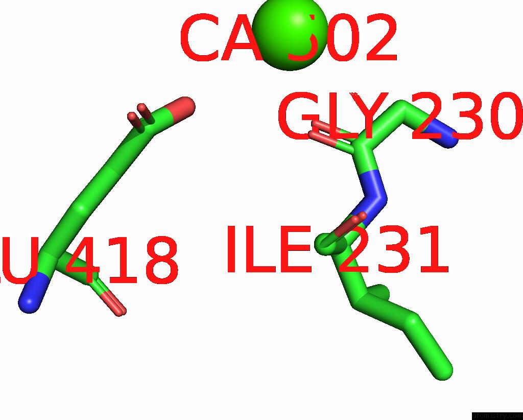
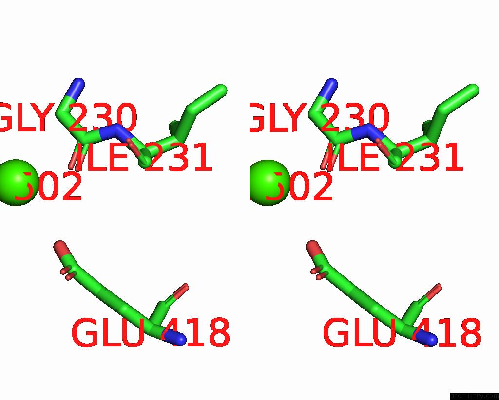
Calcium binding site 1 out of 2 in 7r4j
Go back to
Calcium binding site 1 out
of 2 in the Crystal Structure of Human Mitochondrial Nad Kinase

Mono view

Stereo pair view
Mono view
Stereo pair view
A full contact list of Calcium with other atoms in the Ca binding
site number 1 of Crystal Structure of Human Mitochondrial Nad Kinase within 5.0Å range:
|
Calcium binding site 2 out of 2 in 7r4j
Go back to
Calcium binding site 2 out
of 2 in the Crystal Structure of Human Mitochondrial Nad Kinase
Mono view
Stereo pair view
Mono view
Stereo pair view
A full contact list of Calcium with other atoms in the Ca binding
site number 2 of Crystal Structure of Human Mitochondrial Nad Kinase within 5.0Å range:
|
Reference:
C.Mary,
M.H.Soflaee,
R.Kesavan,
M.Gelin,
H.Brown,
G.Zacharias,
T.P.Mathews,
A.Lemoff,
C.Lionne,
G.Labesse,
G.Hoxhaj.
Crystal Structure of Human NADK2 Reveals A Dimeric Organization and Active Site Occlusion By Lysine Acetylation. Mol.Cell V. 82 3299 2022.
ISSN: ISSN 1097-2765
PubMed: 35868311
DOI: 10.1016/J.MOLCEL.2022.06.026
Page generated: Thu Jul 10 00:41:22 2025
ISSN: ISSN 1097-2765
PubMed: 35868311
DOI: 10.1016/J.MOLCEL.2022.06.026
Last articles
Fe in 2YXOFe in 2YRS
Fe in 2YXC
Fe in 2YNM
Fe in 2YVJ
Fe in 2YP1
Fe in 2YU2
Fe in 2YU1
Fe in 2YQB
Fe in 2YOO