Calcium »
PDB 7qga-7rcc »
7r6b »
Calcium in PDB 7r6b: Crystal Structure of Mutant R43D/L124D/R125A/C273S of L-Asparaginase I From Yersinia Pestis
Enzymatic activity of Crystal Structure of Mutant R43D/L124D/R125A/C273S of L-Asparaginase I From Yersinia Pestis
All present enzymatic activity of Crystal Structure of Mutant R43D/L124D/R125A/C273S of L-Asparaginase I From Yersinia Pestis:
3.5.1.1;
3.5.1.1;
Protein crystallography data
The structure of Crystal Structure of Mutant R43D/L124D/R125A/C273S of L-Asparaginase I From Yersinia Pestis, PDB code: 7r6b
was solved by
P.Strzelczyk,
A.Wlodawer,
J.Lubkowski,
with X-Ray Crystallography technique. A brief refinement statistics is given in the table below:
| Resolution Low / High (Å) | 48.85 / 2.03 |
| Space group | C 2 2 21 |
| Cell size a, b, c (Å), α, β, γ (°) | 100.263, 115.893, 127.548, 90, 90, 90 |
| R / Rfree (%) | 17 / 21.8 |
Calcium Binding Sites:
The binding sites of Calcium atom in the Crystal Structure of Mutant R43D/L124D/R125A/C273S of L-Asparaginase I From Yersinia Pestis
(pdb code 7r6b). This binding sites where shown within
5.0 Angstroms radius around Calcium atom.
In total 2 binding sites of Calcium where determined in the Crystal Structure of Mutant R43D/L124D/R125A/C273S of L-Asparaginase I From Yersinia Pestis, PDB code: 7r6b:
Jump to Calcium binding site number: 1; 2;
In total 2 binding sites of Calcium where determined in the Crystal Structure of Mutant R43D/L124D/R125A/C273S of L-Asparaginase I From Yersinia Pestis, PDB code: 7r6b:
Jump to Calcium binding site number: 1; 2;
Calcium binding site 1 out of 2 in 7r6b
Go back to
Calcium binding site 1 out
of 2 in the Crystal Structure of Mutant R43D/L124D/R125A/C273S of L-Asparaginase I From Yersinia Pestis
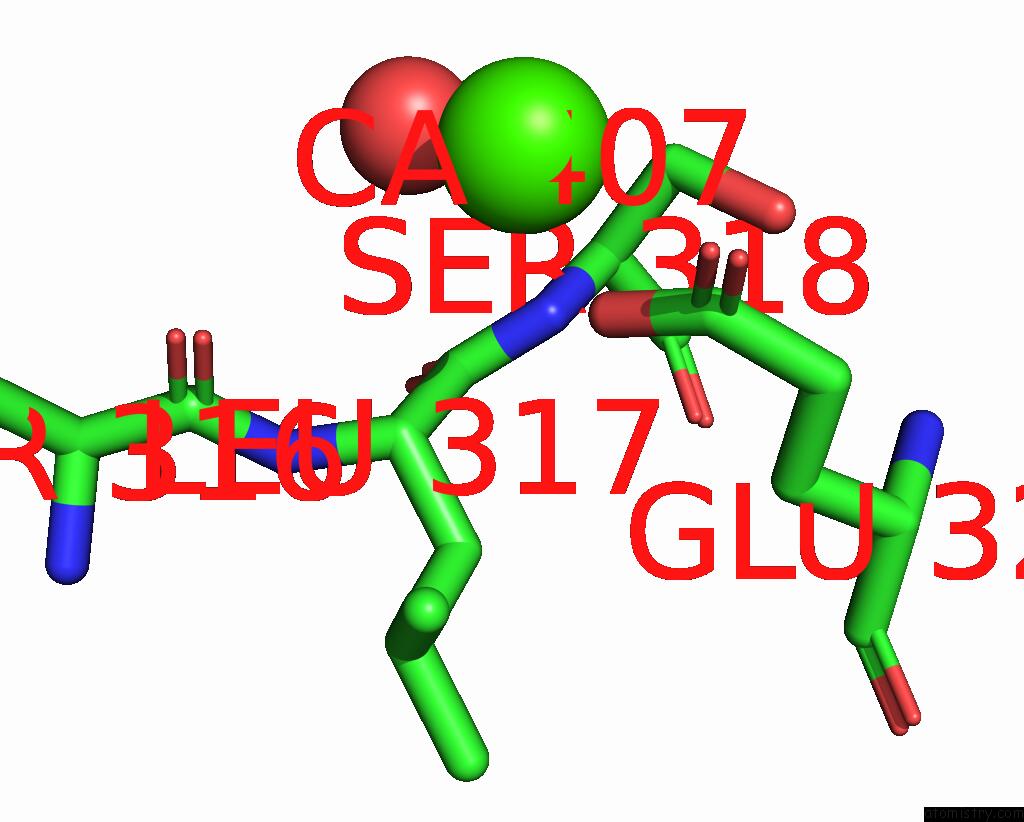
Mono view
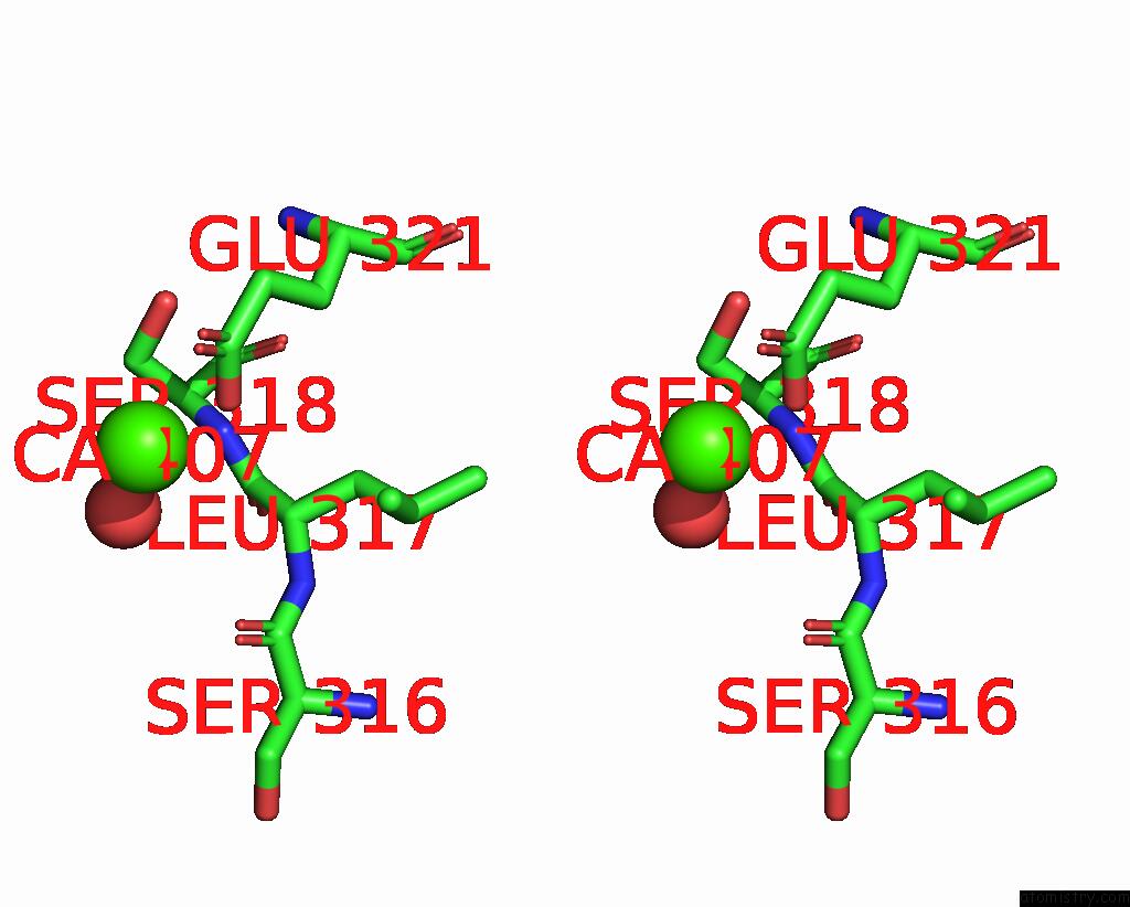
Stereo pair view
Mono view
Stereo pair view
A full contact list of Calcium with other atoms in the Ca binding
site number 1 of Crystal Structure of Mutant R43D/L124D/R125A/C273S of L-Asparaginase I From Yersinia Pestis within 5.0Å range:
|
Calcium binding site 2 out of 2 in 7r6b
Go back to
Calcium binding site 2 out
of 2 in the Crystal Structure of Mutant R43D/L124D/R125A/C273S of L-Asparaginase I From Yersinia Pestis
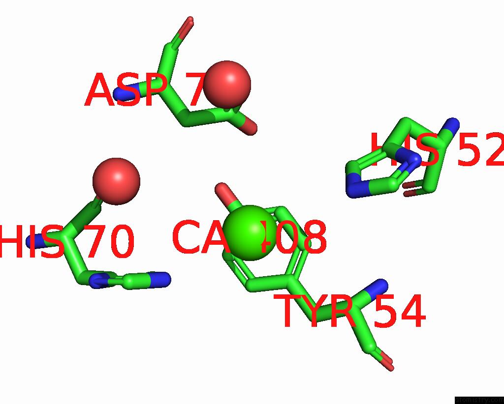
Mono view
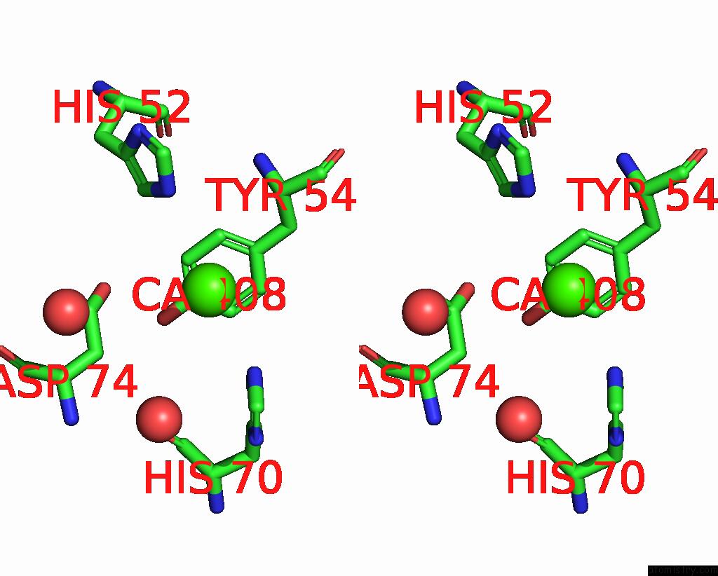
Stereo pair view
Mono view
Stereo pair view
A full contact list of Calcium with other atoms in the Ca binding
site number 2 of Crystal Structure of Mutant R43D/L124D/R125A/C273S of L-Asparaginase I From Yersinia Pestis within 5.0Å range:
|
Reference:
P.Strzelczyk,
D.Zhang,
J.Alexandratos,
G.Piszczek,
A.Wlodawer,
J.Lubkowski.
The Dimeric Form of Bacterial L-Asparaginase Ypai Is Fully Active. Febs J. 2022.
ISSN: ISSN 1742-464X
PubMed: 36152020
DOI: 10.1111/FEBS.16635
Page generated: Thu Jul 10 00:42:04 2025
ISSN: ISSN 1742-464X
PubMed: 36152020
DOI: 10.1111/FEBS.16635
Last articles
Fe in 2YXOFe in 2YRS
Fe in 2YXC
Fe in 2YNM
Fe in 2YVJ
Fe in 2YP1
Fe in 2YU2
Fe in 2YU1
Fe in 2YQB
Fe in 2YOO