Calcium »
PDB 7skm-7tbg »
7sq2 »
Calcium in PDB 7sq2: Reprocessed and Refined Structure of Phospholipase C-Beta and Gq Signaling Complex
Enzymatic activity of Reprocessed and Refined Structure of Phospholipase C-Beta and Gq Signaling Complex
All present enzymatic activity of Reprocessed and Refined Structure of Phospholipase C-Beta and Gq Signaling Complex:
3.1.4.11;
3.1.4.11;
Protein crystallography data
The structure of Reprocessed and Refined Structure of Phospholipase C-Beta and Gq Signaling Complex, PDB code: 7sq2
was solved by
S.T.Endo-Streeter,
J.Sondek,
T.K.Harden,
with X-Ray Crystallography technique. A brief refinement statistics is given in the table below:
| Resolution Low / High (Å) | 40.55 / 2.60 |
| Space group | C 1 2 1 |
| Cell size a, b, c (Å), α, β, γ (°) | 202.837, 90.817, 93.135, 90, 101.19, 90 |
| R / Rfree (%) | 19.9 / 26.3 |
Other elements in 7sq2:
The structure of Reprocessed and Refined Structure of Phospholipase C-Beta and Gq Signaling Complex also contains other interesting chemical elements:
| Aluminium | (Al) | 1 atom |
| Fluorine | (F) | 4 atoms |
| Magnesium | (Mg) | 2 atoms |
Calcium Binding Sites:
The binding sites of Calcium atom in the Reprocessed and Refined Structure of Phospholipase C-Beta and Gq Signaling Complex
(pdb code 7sq2). This binding sites where shown within
5.0 Angstroms radius around Calcium atom.
In total only one binding site of Calcium was determined in the Reprocessed and Refined Structure of Phospholipase C-Beta and Gq Signaling Complex, PDB code: 7sq2:
In total only one binding site of Calcium was determined in the Reprocessed and Refined Structure of Phospholipase C-Beta and Gq Signaling Complex, PDB code: 7sq2:
Calcium binding site 1 out of 1 in 7sq2
Go back to
Calcium binding site 1 out
of 1 in the Reprocessed and Refined Structure of Phospholipase C-Beta and Gq Signaling Complex
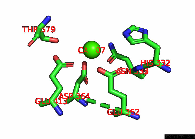
Mono view
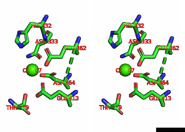
Stereo pair view
Mono view
Stereo pair view
A full contact list of Calcium with other atoms in the Ca binding
site number 1 of Reprocessed and Refined Structure of Phospholipase C-Beta and Gq Signaling Complex within 5.0Å range:
|
Reference:
S.T.Endo-Streeter,
J.Sondek,
T.K.Harden.
Kinetic Scaffolding Mediated By A Phospholipase C-{Beta} and Gq Signaling Complex Science V. 330 974 2010.
ISSN: ISSN 0036-8075
PubMed: 20966218
DOI: 10.1126/SCIENCE.1193438
Page generated: Thu Jul 10 01:06:12 2025
ISSN: ISSN 0036-8075
PubMed: 20966218
DOI: 10.1126/SCIENCE.1193438
Last articles
F in 4JJSF in 4JII
F in 4JDS
F in 4JAV
F in 4JA8
F in 4JBY
F in 4JB4
F in 4JAS
F in 4JA2
F in 4J3J