Calcium »
PDB 7skm-7tbg »
7stt »
Calcium in PDB 7stt: Crystal Structure of Sulfatase From Pedobacter Yulinensis
Protein crystallography data
The structure of Crystal Structure of Sulfatase From Pedobacter Yulinensis, PDB code: 7stt
was solved by
A.O'malley,
C.R.Schlachter,
L.L.Grimes,
J.J.Tomashek,
A.L.Lee,
M.Chruszcz,
with X-Ray Crystallography technique. A brief refinement statistics is given in the table below:
| Resolution Low / High (Å) | 39.37 / 1.60 |
| Space group | P 32 2 1 |
| Cell size a, b, c (Å), α, β, γ (°) | 83.62, 83.62, 116.052, 90, 90, 120 |
| R / Rfree (%) | 14.8 / 17.8 |
Other elements in 7stt:
The structure of Crystal Structure of Sulfatase From Pedobacter Yulinensis also contains other interesting chemical elements:
| Sodium | (Na) | 3 atoms |
| Chlorine | (Cl) | 1 atom |
Calcium Binding Sites:
The binding sites of Calcium atom in the Crystal Structure of Sulfatase From Pedobacter Yulinensis
(pdb code 7stt). This binding sites where shown within
5.0 Angstroms radius around Calcium atom.
In total only one binding site of Calcium was determined in the Crystal Structure of Sulfatase From Pedobacter Yulinensis, PDB code: 7stt:
In total only one binding site of Calcium was determined in the Crystal Structure of Sulfatase From Pedobacter Yulinensis, PDB code: 7stt:
Calcium binding site 1 out of 1 in 7stt
Go back to
Calcium binding site 1 out
of 1 in the Crystal Structure of Sulfatase From Pedobacter Yulinensis
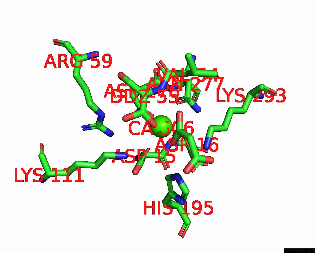
Mono view
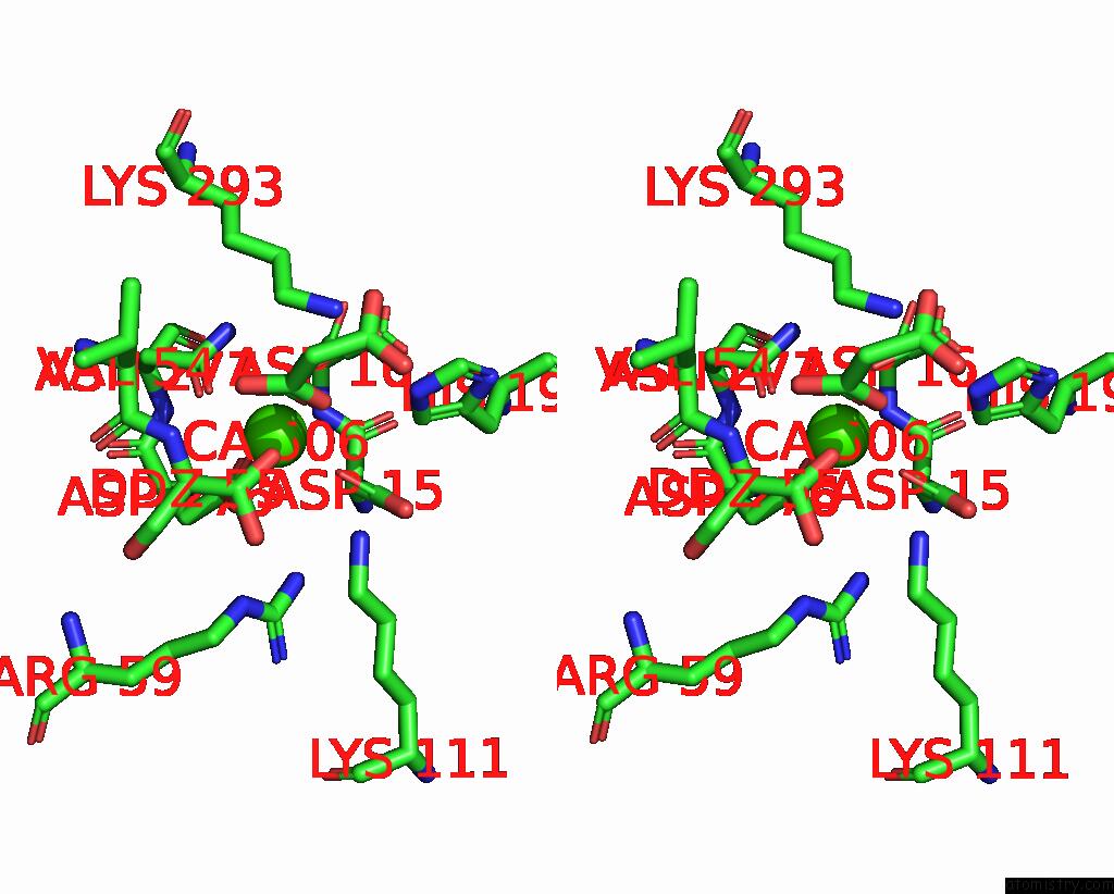
Stereo pair view
Mono view
Stereo pair view
A full contact list of Calcium with other atoms in the Ca binding
site number 1 of Crystal Structure of Sulfatase From Pedobacter Yulinensis within 5.0Å range:
|
Reference:
C.R.Schlachter,
A.O'malley,
L.L.Grimes,
J.J.Tomashek,
M.Chruszcz,
L.A.Lee.
Purification, Characterization, and Structural Studies of A Sulfatase From Pedobacter Yulinensis . Molecules V. 27 2021.
ISSN: ESSN 1420-3049
PubMed: 35011319
DOI: 10.3390/MOLECULES27010087
Page generated: Thu Jul 10 01:07:59 2025
ISSN: ESSN 1420-3049
PubMed: 35011319
DOI: 10.3390/MOLECULES27010087
Last articles
Fe in 2YXOFe in 2YRS
Fe in 2YXC
Fe in 2YNM
Fe in 2YVJ
Fe in 2YP1
Fe in 2YU2
Fe in 2YU1
Fe in 2YQB
Fe in 2YOO