Calcium »
PDB 7skm-7tbg »
7t8d »
Calcium in PDB 7t8d: Myocilin Olf Mutant V449I
Protein crystallography data
The structure of Myocilin Olf Mutant V449I, PDB code: 7t8d
was solved by
H.S.Scelsi,
B.M.Barlow,
R.L.Lieberman,
with X-Ray Crystallography technique. A brief refinement statistics is given in the table below:
| Resolution Low / High (Å) | 35.69 / 1.38 |
| Space group | P 1 21 1 |
| Cell size a, b, c (Å), α, β, γ (°) | 49.408, 50.865, 50.458, 90, 97.01, 90 |
| R / Rfree (%) | 16.3 / 18.4 |
Other elements in 7t8d:
The structure of Myocilin Olf Mutant V449I also contains other interesting chemical elements:
| Sodium | (Na) | 1 atom |
Calcium Binding Sites:
The binding sites of Calcium atom in the Myocilin Olf Mutant V449I
(pdb code 7t8d). This binding sites where shown within
5.0 Angstroms radius around Calcium atom.
In total only one binding site of Calcium was determined in the Myocilin Olf Mutant V449I, PDB code: 7t8d:
In total only one binding site of Calcium was determined in the Myocilin Olf Mutant V449I, PDB code: 7t8d:
Calcium binding site 1 out of 1 in 7t8d
Go back to
Calcium binding site 1 out
of 1 in the Myocilin Olf Mutant V449I
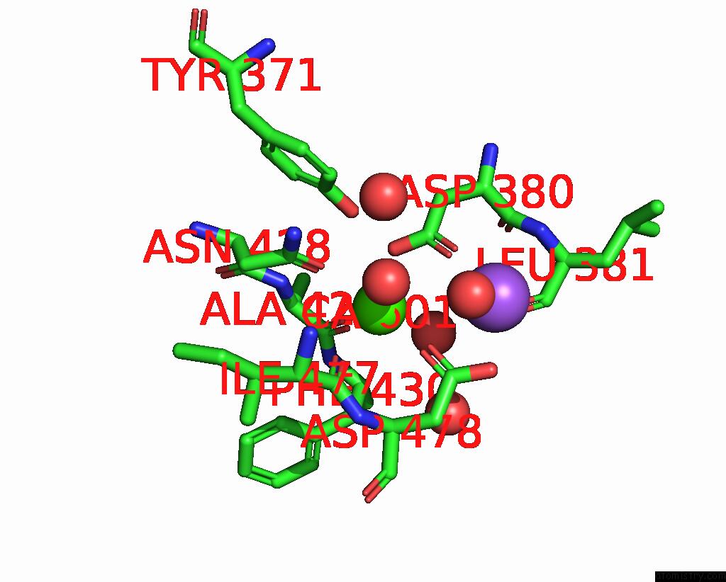
Mono view
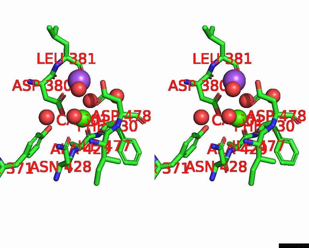
Stereo pair view
Mono view
Stereo pair view
A full contact list of Calcium with other atoms in the Ca binding
site number 1 of Myocilin Olf Mutant V449I within 5.0Å range:
|
Reference:
H.F.Scelsi,
K.R.Hill,
B.M.Barlow,
M.D.Martin,
R.L.Lieberman.
Disambiguation of Benign and Misfolded Glaucoma-Causing Myocilin Variants on the Basis of Protein Thermal Stability. Dis Model Mech 2022.
ISSN: ISSN 1754-8411
PubMed: 36579626
DOI: 10.1242/DMM.049816
Page generated: Thu Jul 10 01:14:50 2025
ISSN: ISSN 1754-8411
PubMed: 36579626
DOI: 10.1242/DMM.049816
Last articles
Cl in 5L52Cl in 5L4H
Cl in 5L4E
Cl in 5L4I
Cl in 5L47
Cl in 5L3H
Cl in 5L3I
Cl in 5L2O
Cl in 5L3X
Cl in 5L39